Malkowski Bogdan, Harat Maciej, Zyromska Agnieszka, Wisniewski Tomasz, Harat Aleksandra, Lopatto Rita, Furtak Jacek
Department of Positron Emission Tomography and Molecular Imaging, Nicolaus Copernicus University, Ludwik Rydygier Collegium Medicum, Bydgoszcz, Poland; Department of Nuclear Medicine, Franciszek Lukaszczyk Oncology Centre, Bydgoszcz, Poland.
Department of Radiotherapy, Franciszek Lukaszczyk Oncology Centre, Bydgoszcz, Poland; Department of Oncology and Brachytherapy, Nicolaus Copernicus University, Ludwik Rydygier Collegium Medicum, Bydgoszcz, Poland.
PLoS One. 2015 Oct 15;10(10):e0140917. doi: 10.1371/journal.pone.0140917. eCollection 2015.
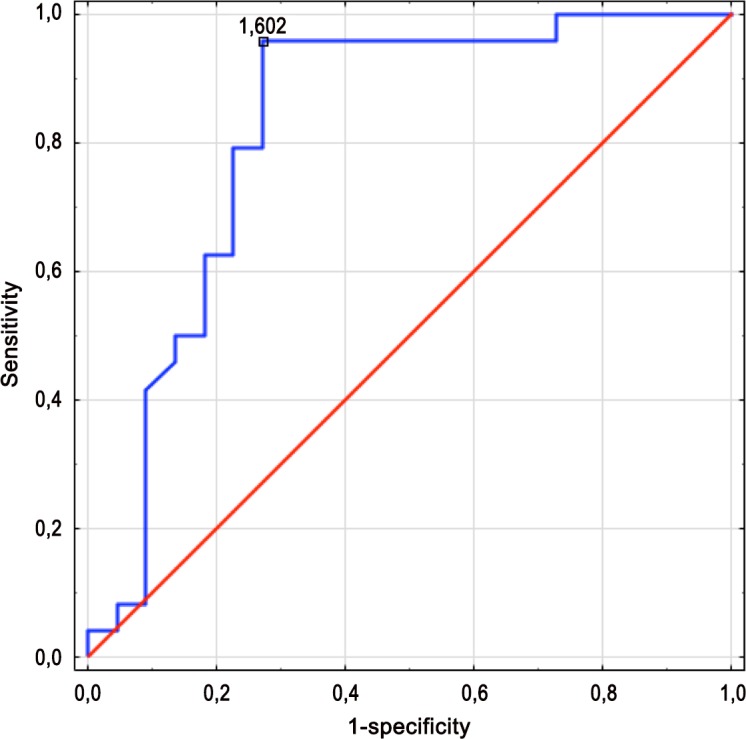
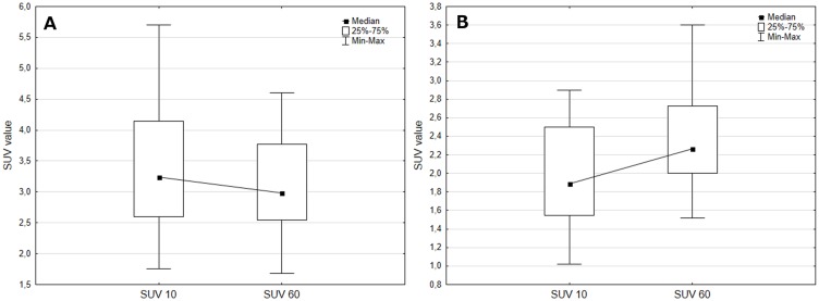
Gliomas are common brain tumours, but obtaining tissue for definitive diagnosis can be difficult. There is, therefore, interest in the use of non-invasive methods to diagnose and grade the disease. Although positron emission tomography (PET) with 18F-fluorethyltyrosine (18F-FET) can be used to differentiate between low-grade (LGG) and high-grade (HGG) gliomas, the optimal parameters to measure and their cut-points have yet to be established. We therefore assessed the value of single and dual time-point acquisition of 18F-FET PET parameters to differentiate between primary LGGs (n = 22) and HGGs (n = 24). PET examination was considered positive for glioma if the metabolic activity was 1.6-times higher than that of background (contralateral) brain, and maximum tissue-brain ratios (TBRmax) were calculated 10 and 60 min after isotope administration with their sums and differences calculated from individual time-point values. Using a threshold-based method, the overall sensitivity of PET was 97%. Several analysed parameters were significantly different between LGGs and HGGs. However, in a receiver operating characteristics analysis, TBR sum had the best diagnostic accuracy of 87% and sensitivity, specificity, and positive and negative predictive values of 100%, 72.7%, 80%, and 100%, respectively. 18F-FET PET is valuable for the non-invasive determination of glioma grade, especially when dual time-point metrics are used. TBR sum shows the greatest accuracy, sensitivity, and negative predictive value for tumour grade differentiation and is a simple method to implement. However, the cut-off may differ between institutions and calibration strategies would be useful.
胶质瘤是常见的脑肿瘤,但获取用于明确诊断的组织可能很困难。因此,人们对使用非侵入性方法来诊断和分级该疾病很感兴趣。尽管正电子发射断层扫描(PET)联合18F-氟乙基酪氨酸(18F-FET)可用于区分低级别(LGG)和高级别(HGG)胶质瘤,但测量的最佳参数及其切点尚未确定。因此,我们评估了18F-FET PET参数单时间点和双时间点采集对区分原发性LGG(n = 22)和HGG(n = 24)的价值。如果代谢活性比背景(对侧)脑高1.6倍,则PET检查被认为对胶质瘤呈阳性,并在同位素给药后10分钟和60分钟计算最大组织-脑比率(TBRmax),并根据各个时间点的值计算其总和与差值。使用基于阈值的方法,PET的总体敏感性为97%。LGG和HGG之间的几个分析参数有显著差异。然而,在受试者工作特征分析中,TBR总和的诊断准确性最高,为87%,敏感性、特异性、阳性和阴性预测值分别为100%、72.7%、80%和100%。18F-FET PET对于非侵入性确定胶质瘤分级很有价值,尤其是在使用双时间点指标时。TBR总和在肿瘤分级区分方面显示出最高的准确性、敏感性和阴性预测值,并且是一种易于实施的简单方法。然而,不同机构的临界值可能不同,校准策略将很有用。