Kusuma Gina D, Menicanin Danijela, Gronthos Stan, Manuelpillai Ursula, Abumaree Mohamed H, Pertile Mark D, Brennecke Shaun P, Kalionis Bill
Department of Obstetrics and Gynaecology, Royal Women's Hospital, The University of Melbourne, Parkville, Victoria, Australia; Pregnancy Research Centre, Department of Perinatal Medicine, Royal Women's Hospital, Parkville, Victoria, Australia.
Mesenchymal Stem Cell Laboratory, Faculty of Health Sciences, School of Medical Sciences, University of Adelaide, Adelaide, Australia; Colgate Australian Clinical Dental Research Centre, School of Dentistry, University of Adelaide, Adelaide, Australia.
PLoS One. 2015 Oct 20;10(10):e0141246. doi: 10.1371/journal.pone.0141246. eCollection 2015.
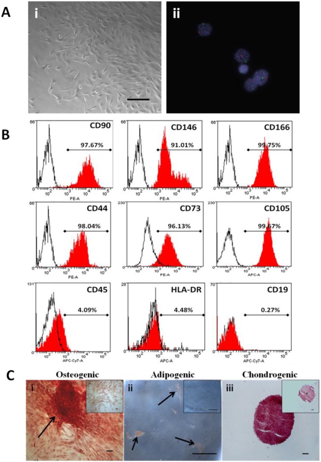
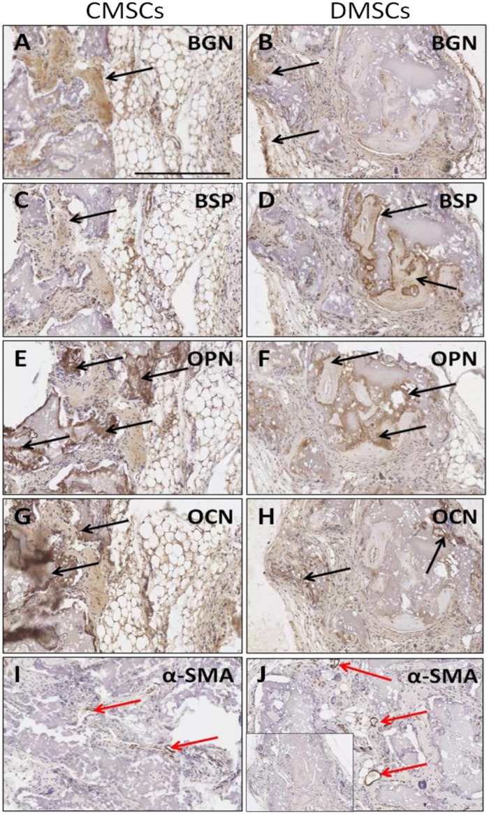
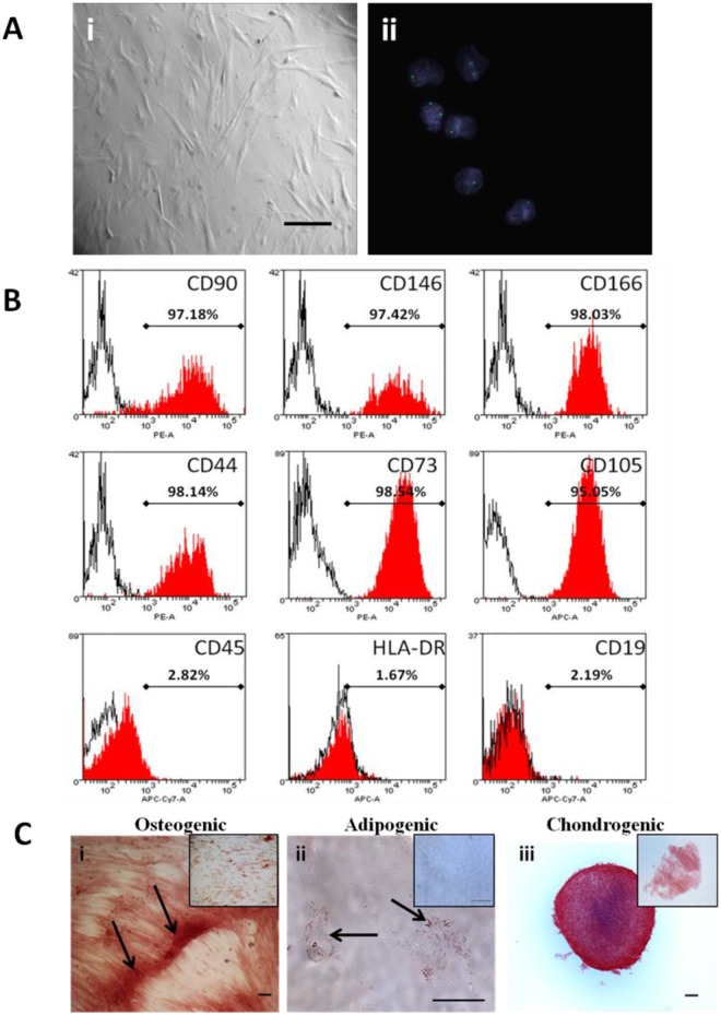
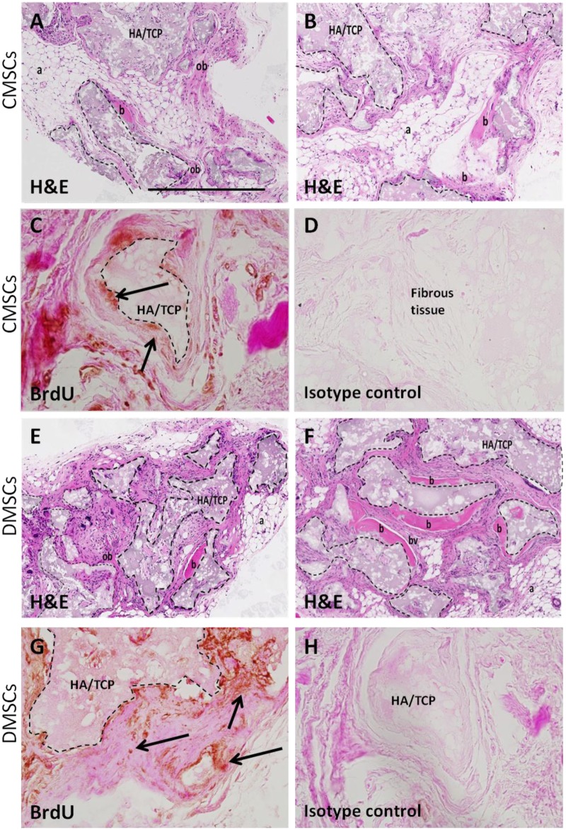
Mesenchymal stem cells (MSCs) are one of the most attractive cell types for cell-based bone tissue repair applications. Fetal-derived MSCs and maternal-derived MSCs have been isolated from chorionic villi of human term placenta and the decidua basalis attached to the placenta following delivery, respectively. Chorionic-derived MSCs (CMSCs) and decidua-derived MSCs (DMSCs) generated in this study met the MSCs criteria set by International Society of Cellular Therapy. These criteria include: (i) adherence to plastic; (ii) >90% expression of CD73, CD105, CD90, CD146, CD44 and CD166 combined with <5% expression of CD45, CD19 and HLA-DR; and (iii) ability to differentiate into osteogenic, adipogenic, and chondrogenic lineages. In vivo subcutaneous implantation into SCID mice showed that both bromo-deoxyuridine (BrdU)-labelled CMSCs and DMSCs when implanted together with hydroxyapatite/tricalcium phosphate particles were capable of forming ectopic bone at 8-weeks post-transplantation. Histological assessment showed expression of bone markers, osteopontin (OPN), osteocalcin (OCN), biglycan (BGN), bone sialoprotein (BSP), and also a marker of vasculature, alpha-smooth muscle actin (α-SMA). This study provides evidence to support CMSCs and DMSCs as cellular candidates with potent bone forming capacity.
间充质干细胞(MSCs)是基于细胞的骨组织修复应用中最具吸引力的细胞类型之一。胎儿来源的间充质干细胞和母体来源的间充质干细胞分别从足月人胎盘的绒毛膜绒毛和分娩后附着于胎盘的基蜕膜中分离得到。本研究中产生的绒毛膜来源的间充质干细胞(CMSCs)和蜕膜来源的间充质干细胞(DMSCs)符合国际细胞治疗协会设定的间充质干细胞标准。这些标准包括:(i)贴壁于塑料培养皿;(ii)CD73、CD105、CD90、CD146、CD44和CD166的表达>90%,同时CD45、CD19和HLA-DR的表达<5%;(iii)能够分化为成骨、成脂和成软骨谱系。在严重联合免疫缺陷(SCID)小鼠体内皮下植入实验表明,与羟基磷灰石/磷酸三钙颗粒一起植入时,溴脱氧尿苷(BrdU)标记的CMSCs和DMSCs在移植后8周均能够形成异位骨。组织学评估显示骨标志物骨桥蛋白(OPN)、骨钙素(OCN)、双糖链蛋白聚糖(BGN)、骨唾液蛋白(BSP)以及血管标志物α-平滑肌肌动蛋白(α-SMA)均有表达。本研究提供了证据支持CMSCs和DMSCs作为具有强大骨形成能力的细胞候选物。