• 文献检索
  • 文档翻译
  • 深度研究
  • 学术资讯
  • Suppr Zotero 插件Zotero 插件
  • 邀请有礼
  • 套餐&价格
  • 历史记录
应用&插件
Suppr Zotero 插件Zotero 插件浏览器插件Mac 客户端Windows 客户端微信小程序
定价
高级版会员购买积分包购买API积分包
服务
文献检索文档翻译深度研究API 文档MCP 服务
关于我们
关于 Suppr公司介绍联系我们用户协议隐私条款
关注我们

Suppr 超能文献

核心技术专利:CN118964589B侵权必究
粤ICP备2023148730 号-1Suppr @ 2026

文献检索

告别复杂PubMed语法,用中文像聊天一样搜索,搜遍4000万医学文献。AI智能推荐,让科研检索更轻松。

立即免费搜索

文件翻译

保留排版,准确专业,支持PDF/Word/PPT等文件格式,支持 12+语言互译。

免费翻译文档

深度研究

AI帮你快速写综述,25分钟生成高质量综述,智能提取关键信息,辅助科研写作。

立即免费体验

使用纳米颗粒造影剂对小鼠模型中的肝细胞癌进行X射线散射成像。

X-ray Scatter Imaging of Hepatocellular Carcinoma in a Mouse Model Using Nanoparticle Contrast Agents.

作者信息

Rand Danielle, Derdak Zoltan, Carlson Rolf, Wands Jack R, Rose-Petruck Christoph

机构信息

Department of Chemistry, Brown University. 324 Brook Street, Providence, Rhode Island 02912 (USA).

The Liver Research Center, Rhode Island Hospital and Warren Alpert Medical School of Brown University. 55 Claverick Street, Providence, Rhode Island 02903 (USA).

出版信息

Sci Rep. 2015 Oct 29;5:15673. doi: 10.1038/srep15673.

DOI:10.1038/srep15673
PMID:26511147
原文链接:https://pmc.ncbi.nlm.nih.gov/articles/PMC4625152/
Abstract

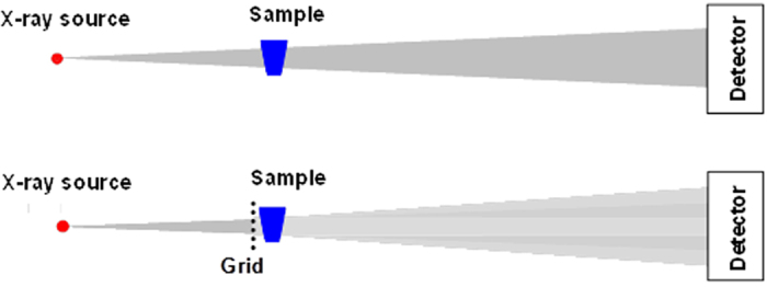
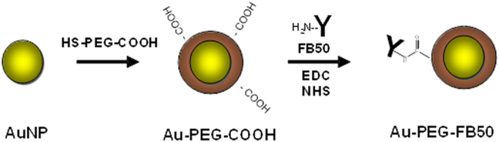
Hepatocellular carcinoma (HCC) is one of the most common malignant tumors worldwide and is almost uniformly fatal. Current methods of detection include ultrasound examination and imaging by CT scan or MRI; however, these techniques are problematic in terms of sensitivity and specificity, and the detection of early tumors (<1 cm diameter) has proven elusive. Better, more specific, and more sensitive detection methods are therefore urgently needed. Here we discuss the application of a newly developed x-ray imaging technique called Spatial Frequency Heterodyne Imaging (SFHI) for the early detection of HCC. SFHI uses x-rays scattered by an object to form an image and is more sensitive than conventional absorption-based x-radiography. We show that tissues labeled in vivo with gold nanoparticle contrast agents can be detected using SFHI. We also demonstrate that directed targeting and SFHI of HCC tumors in a mouse model is possible through the use of HCC-specific antibodies. The enhanced sensitivity of SFHI relative to currently available techniques enables the x-ray imaging of tumors that are just a few millimeters in diameter and substantially reduces the amount of nanoparticle contrast agent required for intravenous injection relative to absorption-based x-ray imaging.

摘要

肝细胞癌(HCC)是全球最常见的恶性肿瘤之一,几乎无一例外都是致命的。目前的检测方法包括超声检查以及CT扫描或MRI成像;然而,这些技术在灵敏度和特异性方面存在问题,而且事实证明,早期肿瘤(直径<1厘米)的检测很难实现。因此,迫切需要更好、更特异且更灵敏的检测方法。在此,我们讨论一种新开发的名为空间频率外差成像(SFHI)的X射线成像技术在HCC早期检测中的应用。SFHI利用物体散射的X射线形成图像,比传统的基于吸收的X射线摄影更灵敏。我们表明,使用SFHI可以检测体内用金纳米颗粒造影剂标记的组织。我们还证明,通过使用HCC特异性抗体,在小鼠模型中对HCC肿瘤进行定向靶向和SFHI检测是可行的。相对于目前可用的技术,SFHI更高的灵敏度能够对直径仅几毫米的肿瘤进行X射线成像,并且相对于基于吸收的X射线成像,大大减少了静脉注射所需的纳米颗粒造影剂的用量。

https://cdn.ncbi.nlm.nih.gov/pmc/blobs/9c43/4625152/2237584724ee/srep15673-f10.jpg
https://cdn.ncbi.nlm.nih.gov/pmc/blobs/9c43/4625152/2e82f6c143f6/srep15673-f1.jpg
https://cdn.ncbi.nlm.nih.gov/pmc/blobs/9c43/4625152/61f5c19865f7/srep15673-f2.jpg
https://cdn.ncbi.nlm.nih.gov/pmc/blobs/9c43/4625152/b9fc48b669db/srep15673-f3.jpg
https://cdn.ncbi.nlm.nih.gov/pmc/blobs/9c43/4625152/92310b3bb620/srep15673-f4.jpg
https://cdn.ncbi.nlm.nih.gov/pmc/blobs/9c43/4625152/58558a23d04b/srep15673-f5.jpg
https://cdn.ncbi.nlm.nih.gov/pmc/blobs/9c43/4625152/71c496131ce8/srep15673-f6.jpg
https://cdn.ncbi.nlm.nih.gov/pmc/blobs/9c43/4625152/2213353745b9/srep15673-f7.jpg
https://cdn.ncbi.nlm.nih.gov/pmc/blobs/9c43/4625152/318da20593e6/srep15673-f8.jpg
https://cdn.ncbi.nlm.nih.gov/pmc/blobs/9c43/4625152/de1aa21afdab/srep15673-f9.jpg
https://cdn.ncbi.nlm.nih.gov/pmc/blobs/9c43/4625152/2237584724ee/srep15673-f10.jpg

相似文献

1
X-ray Scatter Imaging of Hepatocellular Carcinoma in a Mouse Model Using Nanoparticle Contrast Agents.使用纳米颗粒造影剂对小鼠模型中的肝细胞癌进行X射线散射成像。
Sci Rep. 2015 Oct 29;5:15673. doi: 10.1038/srep15673.
2
A highly sensitive x-ray imaging modality for hepatocellular carcinoma detection in vitro.一种用于体外检测肝细胞癌的高灵敏度X射线成像模态。
Phys Med Biol. 2015 Jan 21;60(2):769-84. doi: 10.1088/0031-9155/60/2/769. Epub 2015 Jan 5.
3
X-ray spatial frequency heterodyne imaging of protein-based nanobubble contrast agents.基于蛋白质的纳米气泡造影剂的X射线空间频率外差成像
Opt Express. 2014 Sep 22;22(19):23290-8. doi: 10.1364/OE.22.023290.
4
Time course of contrast enhancement by micro-CT with dedicated contrast agents in normal mice and mice with hepatocellular carcinoma: comparison of one iodinated and two nanoparticle-based agents.使用专用造影剂通过微型计算机断层扫描(micro-CT)对正常小鼠和肝细胞癌小鼠进行对比增强的时间进程:一种碘化剂和两种基于纳米颗粒的造影剂的比较
Acad Radiol. 2015 Feb;22(2):169-78. doi: 10.1016/j.acra.2014.07.022. Epub 2014 Oct 1.
5
Biodegradable human serum albumin nanoparticles as contrast agents for the detection of hepatocellular carcinoma by magnetic resonance imaging.可生物降解的人血清白蛋白纳米颗粒作为磁共振成像检测肝细胞癌的对比剂。
Eur J Pharm Biopharm. 2014 May;87(1):132-41. doi: 10.1016/j.ejpb.2013.12.010. Epub 2013 Dec 21.
6
Characterization of malignant focal liver lesions with contrast-enhanced 40 MHz ultrasound imaging in hepatitis B virus X transgenic mice: a feasibility study.在乙型肝炎病毒X转基因小鼠中用40兆赫超声造影成像对恶性局灶性肝病变进行特征分析:一项可行性研究。
Ultrason Imaging. 2008 Oct;30(4):203-16. doi: 10.1177/016173460803000402.
7
Detection of hepatocellular carcinoma in transgenic mice by Gd-DTPA- and rhodamine 123-conjugated human serum albumin nanoparticles in T1 magnetic resonance imaging.利用 Gd-DTPA 和 rhodamine 123 偶联人血清白蛋白纳米粒在 T1 磁共振成像中检测转基因小鼠肝癌。
J Control Release. 2015 Feb 10;199:63-71. doi: 10.1016/j.jconrel.2014.11.023. Epub 2014 Dec 10.
8
Is 3-Tesla Gd-EOB-DTPA-enhanced MRI with diffusion-weighted imaging superior to 64-slice contrast-enhanced CT for the diagnosis of hepatocellular carcinoma?3 特斯拉钆塞酸二钠增强磁共振成像联合弥散加权成像在肝细胞癌诊断中是否优于 64 层螺旋 CT 增强扫描?
PLoS One. 2014 Nov 6;9(11):e111935. doi: 10.1371/journal.pone.0111935. eCollection 2014.
9
Predictive Value of Conventional Ultrasound and Contrast-Enhanced Ultrasound in Early Recurrence of Hepatocellular Carcinoma after Surgical Resection.常规超声及超声造影对肝细胞癌手术切除后早期复发的预测价值
Ultrasound Med Biol. 2016 May;42(5):1042-8. doi: 10.1016/j.ultrasmedbio.2015.12.010. Epub 2016 Jan 21.
10
Nanomaterials for X-ray imaging: gold nanoparticle enhancement of X-ray scatter imaging of hepatocellular carcinoma.用于 X 射线成像的纳米材料:金纳米颗粒增强肝细胞癌的 X 射线散射成像。
Nano Lett. 2011 Jul 13;11(7):2678-83. doi: 10.1021/nl200858y. Epub 2011 Jun 6.

本文引用的文献

1
A highly sensitive x-ray imaging modality for hepatocellular carcinoma detection in vitro.一种用于体外检测肝细胞癌的高灵敏度X射线成像模态。
Phys Med Biol. 2015 Jan 21;60(2):769-84. doi: 10.1088/0031-9155/60/2/769. Epub 2015 Jan 5.
2
X-ray spatial frequency heterodyne imaging of protein-based nanobubble contrast agents.基于蛋白质的纳米气泡造影剂的X射线空间频率外差成像
Opt Express. 2014 Sep 22;22(19):23290-8. doi: 10.1364/OE.22.023290.
3
Kupffer cells in the liver.肝脏中的库普弗细胞。
Compr Physiol. 2013 Apr;3(2):785-97. doi: 10.1002/cphy.c120026.
4
Functional imaging techniques in hepatocellular carcinoma.肝细胞癌的功能成像技术。
Eur J Nucl Med Mol Imaging. 2012 Jun;39(6):1070-9. doi: 10.1007/s00259-012-2096-x. Epub 2012 Mar 21.
5
Ultrasonography, computed tomography and magnetic resonance imaging of hepatocellular carcinoma: toward improved treatment decisions.肝细胞癌的超声、计算机断层扫描和磁共振成像:走向更好的治疗决策。
Oncology. 2011;81 Suppl 1:86-99. doi: 10.1159/000333267. Epub 2011 Dec 22.
6
Diagnostic imaging of hepatocellular carcinoma: recent progress.肝细胞癌的诊断影像学:最新进展。
Oncology. 2011;81 Suppl 1:73-85. doi: 10.1159/000333265. Epub 2011 Dec 22.
7
The first analysis and clinical evaluation of native breast tissue using differential phase-contrast mammography.利用差分相衬乳腺摄影术对原生乳腺组织进行的首次分析和临床评估。
Invest Radiol. 2011 Dec;46(12):801-6. doi: 10.1097/RLI.0b013e31822a585f.
8
X-ray spatial harmonic imaging of phase objects.X 射线相位物体的空间谐波成象。
Opt Lett. 2011 Jun 15;36(12):2209-11. doi: 10.1364/OL.36.002209.
9
Nanomaterials for X-ray imaging: gold nanoparticle enhancement of X-ray scatter imaging of hepatocellular carcinoma.用于 X 射线成像的纳米材料:金纳米颗粒增强肝细胞癌的 X 射线散射成像。
Nano Lett. 2011 Jul 13;11(7):2678-83. doi: 10.1021/nl200858y. Epub 2011 Jun 6.
10
Global cancer statistics.全球癌症统计数据。
CA Cancer J Clin. 2011 Mar-Apr;61(2):69-90. doi: 10.3322/caac.20107. Epub 2011 Feb 4.