DaMata Jarina Pena, Mendes Bárbara Pinheiro, Maciel-Lima Kátia, Menezes Cristiane Alves Silva, Dutra Walderez Ornelas, Sousa Lirlândia Pires, Horta Maria Fátima
Departamento de Bioquímica e Imunologia, Instituto de Ciências Biológicas, Universidade Federal de Minas Gerais, Belo Horizonte, MG, Brazil.
Departamento de Morfologia, Instituto de Ciências Biológicas, Universidade Federal de Minas Gerais, Belo Horizonte, MG, Brazil.
PLoS One. 2015 Oct 29;10(10):e0141196. doi: 10.1371/journal.pone.0141196. eCollection 2015.
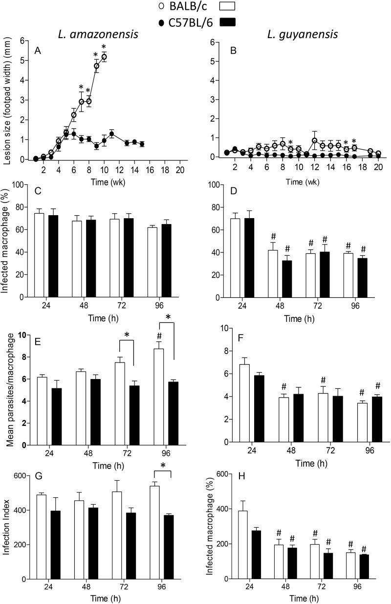
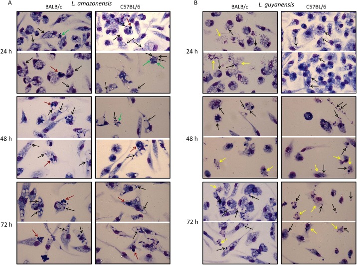
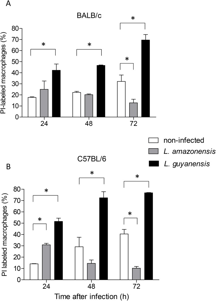
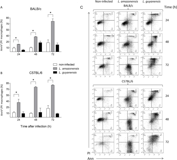
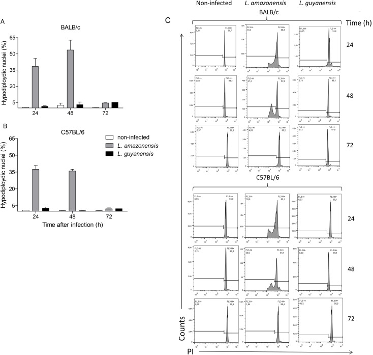
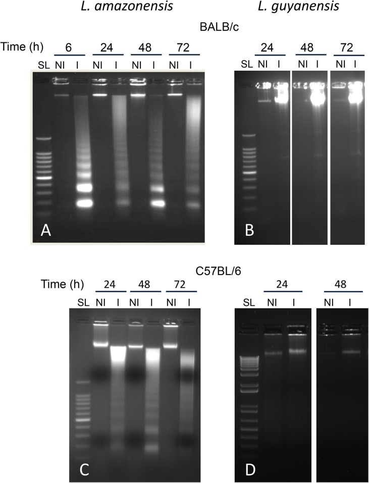
Leishmania is an intracellular parasite in vertebrate hosts, including man. During infection, amastigotes replicate inside macrophages and are transmitted to healthy cells, leading to amplification of the infection. Although transfer of amastigotes from infected to healthy cells is a crucial step that may shape the outcome of the infection, it is not fully understood. Here we compare L. amazonensis and L. guyanensis infection in C57BL/6 and BALB/c mice and investigate the fate of macrophages when infected with these species of Leishmania in vitro. As previously shown, infection of mice results in distinct outcomes: L. amazonensis causes a chronic infection in both strains of mice (although milder in C57BL/6), whereas L. guyanensis does not cause them disease. In vitro, infection is persistent in L. amazonensis-infected macrophages whereas L. guyanensis growth is controlled by host cells from both strains of mice. We demonstrate that, in vitro, L. amazonensis induces apoptosis of both C57BL/6 and BALB/c macrophages, characterized by PS exposure, DNA cleavage into nucleosomal size fragments, and consequent hypodiploidy. None of these signs were seen in macrophages infected with L. guyanensis, which seem to die through necrosis, as indicated by increased PI-, but not Annexin V-, positive cells. L. amazonensis-induced macrophage apoptosis was associated to activation of caspases-3, -8 and -9 in both strains of mice. Considering these two species of Leishmania and strains of mice, macrophage apoptosis, induced at the initial moments of infection, correlates with chronic infection, regardless of its severity. We present evidence suggestive that macrophages phagocytize L. amazonensis-infected cells, which has not been verified so far. The ingestion of apoptotic infected macrophages by healthy macrophages could be a way of amastigote spreading, leading to the establishment of infection.
利什曼原虫是包括人类在内的脊椎动物宿主中的一种细胞内寄生虫。在感染过程中,无鞭毛体在巨噬细胞内复制并传播到健康细胞,导致感染扩大。尽管无鞭毛体从受感染细胞转移到健康细胞是一个可能决定感染结果的关键步骤,但人们对此尚未完全了解。在这里,我们比较了亚马逊利什曼原虫和圭亚那利什曼原虫在C57BL/6和BALB/c小鼠中的感染情况,并研究了在体外感染这些利什曼原虫物种时巨噬细胞的命运。如先前所示,小鼠感染会导致不同的结果:亚马逊利什曼原虫在两种小鼠品系中都会引起慢性感染(尽管在C57BL/6中较轻),而圭亚那利什曼原虫不会使它们患病。在体外,亚马逊利什曼原虫感染的巨噬细胞中感染持续存在,而圭亚那利什曼原虫的生长受到两种小鼠品系宿主细胞的控制。我们证明,在体外,亚马逊利什曼原虫诱导C57BL/6和BALB/c巨噬细胞凋亡,其特征为磷脂酰丝氨酸暴露、DNA裂解成核小体大小的片段以及随之而来的亚二倍体。在感染圭亚那利什曼原虫的巨噬细胞中未观察到这些迹象,这些细胞似乎通过坏死死亡,这由碘化丙啶(PI)阳性细胞增加所表明,但膜联蛋白V(Annexin V)阳性细胞未增加。亚马逊利什曼原虫诱导的巨噬细胞凋亡与两种小鼠品系中半胱天冬酶-3、-8和-9的激活有关。考虑到这两种利什曼原虫物种和小鼠品系,在感染初期诱导的巨噬细胞凋亡与慢性感染相关,无论其严重程度如何。我们提供的证据表明巨噬细胞吞噬了感染亚马逊利什曼原虫的细胞,这一点迄今尚未得到证实。健康巨噬细胞摄取凋亡的受感染巨噬细胞可能是无鞭毛体传播的一种方式,从而导致感染的建立。