• 文献检索
  • 文档翻译
  • 深度研究
  • 学术资讯
  • Suppr Zotero 插件Zotero 插件
  • 邀请有礼
  • 套餐&价格
  • 历史记录
应用&插件
Suppr Zotero 插件Zotero 插件浏览器插件Mac 客户端Windows 客户端微信小程序
定价
高级版会员购买积分包购买API积分包
服务
文献检索文档翻译深度研究API 文档MCP 服务
关于我们
关于 Suppr公司介绍联系我们用户协议隐私条款
关注我们

Suppr 超能文献

核心技术专利:CN118964589B侵权必究
粤ICP备2023148730 号-1Suppr @ 2026

文献检索

告别复杂PubMed语法,用中文像聊天一样搜索,搜遍4000万医学文献。AI智能推荐,让科研检索更轻松。

立即免费搜索

文件翻译

保留排版,准确专业,支持PDF/Word/PPT等文件格式,支持 12+语言互译。

免费翻译文档

深度研究

AI帮你快速写综述,25分钟生成高质量综述,智能提取关键信息,辅助科研写作。

立即免费体验

评估定义ΔFA%截断点以区分四种主要类型的肿瘤周围白质束受累情况的可能性。

Evaluating the Possibility of Defining Cut-Off Points for ΔFA% in Order to Differentiate Four Major Types of Peri-Tumoral White Matter Tract Involvement.

作者信息

Deilami Tourisa, Hadizadeh Kharrazi Homayoun, Seddighi Amir Saied, Tanzifi Parin, Tayebivaljouzi Reza, Zamani Fatemeh, Chavoshzadeh Tafti Atefeh

机构信息

Department of Radiology, Tehran University of Medical Sciences, Tehran, Iran.

Department of Neurosurgery, Shahid Beheshti University of Medical Sciences, Tehran, Iran.

出版信息

Iran J Radiol. 2015 Jul 22;12(3):e9567. doi: 10.5812/iranjradiol.9567v2. eCollection 2015 Jul.

DOI:10.5812/iranjradiol.9567v2
PMID:26528388
原文链接:https://pmc.ncbi.nlm.nih.gov/articles/PMC4623775/
Abstract

BACKGROUND

Diffusion tensor imaging (DTI) and its different scalar values such as fractional anisotropy (FA) have recently been used for evaluation of peri-tumoral white matter (WM) involvement to help define safer surgical excision margins.

OBJECTIVES

The purpose of this study is to evaluate the possibility of defining diagnostic cut-off points for differentiating four major types of peri-tumoral WM involvement using FA.

PATIENTS AND METHODS

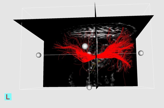
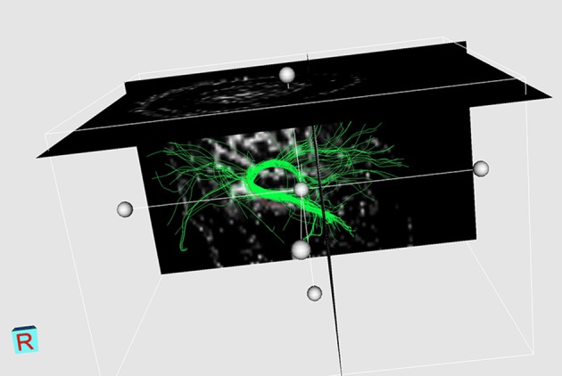
DTI was performed in 12 patients with high presumption of having brain tumors, on a 1.5 T MRI scanner. DTI data was processed by MedINRIA software. Two-hundred region of interests (ROI) were evaluated: 100 in the lesion zone and the rest in the normal WM in the contralateral hemisphere. FA value related to each ROI was measured, and the percentage of FA decrement (ΔFAs%) was calculated.

RESULTS

Of the 100 ROIs on the lesion side, 74 were related to high-grade lesions, 23 to low-grade ones, and three to "gliosis". There were 54 "infiltrated", 22 "displaced", 15 "disrupted", and 9 "edematous" tracts. The major type of fiber involvement, both in low-grade and high-grade tumors was "infiltrated, whereas "edematous" fibers comprised the minority. ΔFA% was more than -35 for "displaced" and "edematous" fibers, and less than -35 for the majority of "disrupted" ones, but "infiltrated" fibers had scattered distribution. Mean ΔFA% was the least for "disrupted", followed by "infiltrated", "edematous" and "displaced" parts.

CONCLUSION

Introducing definite diagnostic cut-points was not possible, due to overlap. Based on the fact that "disruption" is the most aggressive process, a sensitivity analysis was carried out for "disrupted" fibers for several presumptive cut-off points.

摘要

背景

扩散张量成像(DTI)及其不同的标量值,如分数各向异性(FA),最近已被用于评估肿瘤周围白质(WM)受累情况,以帮助确定更安全的手术切除边缘。

目的

本研究的目的是评估使用FA定义诊断分界点以区分四种主要类型的肿瘤周围WM受累情况的可能性。

患者和方法

对12例高度疑似患有脑肿瘤的患者在1.5T MRI扫描仪上进行DTI检查。DTI数据由MedINRIA软件处理。评估了200个感兴趣区域(ROI):100个在病变区域,其余在对侧半球的正常WM中。测量与每个ROI相关的FA值,并计算FA降低百分比(ΔFAs%)。

结果

病变侧的100个ROI中,74个与高级别病变相关,23个与低级别病变相关,3个与“胶质增生”相关。有54条“浸润性”、22条“移位性”、15条“中断性”和9条“水肿性”纤维束。在低级别和高级别肿瘤中,纤维受累的主要类型均为“浸润性”,而“水肿性

https://cdn.ncbi.nlm.nih.gov/pmc/blobs/9f80/4623775/c99517a6a51c/iranjradiol-12-03-9567-g003.jpg
https://cdn.ncbi.nlm.nih.gov/pmc/blobs/9f80/4623775/457058ae43cd/iranjradiol-12-03-9567-g001.jpg
https://cdn.ncbi.nlm.nih.gov/pmc/blobs/9f80/4623775/6f598e8e3ba8/iranjradiol-12-03-9567-g002.jpg
https://cdn.ncbi.nlm.nih.gov/pmc/blobs/9f80/4623775/c99517a6a51c/iranjradiol-12-03-9567-g003.jpg
https://cdn.ncbi.nlm.nih.gov/pmc/blobs/9f80/4623775/457058ae43cd/iranjradiol-12-03-9567-g001.jpg
https://cdn.ncbi.nlm.nih.gov/pmc/blobs/9f80/4623775/6f598e8e3ba8/iranjradiol-12-03-9567-g002.jpg
https://cdn.ncbi.nlm.nih.gov/pmc/blobs/9f80/4623775/c99517a6a51c/iranjradiol-12-03-9567-g003.jpg

相似文献

1
Evaluating the Possibility of Defining Cut-Off Points for ΔFA% in Order to Differentiate Four Major Types of Peri-Tumoral White Matter Tract Involvement.评估定义ΔFA%截断点以区分四种主要类型的肿瘤周围白质束受累情况的可能性。
Iran J Radiol. 2015 Jul 22;12(3):e9567. doi: 10.5812/iranjradiol.9567v2. eCollection 2015 Jul.
2
White Matter tract involvement in brain tumors: a diffusion tensor imaging analysis.脑肿瘤中的白质纤维束受累:一项扩散张量成像分析
Surg Neurol. 2009 Nov;72(5):464-9; discussion 469. doi: 10.1016/j.surneu.2009.05.008. Epub 2009 Jul 15.
3
The role of diffusion tensor imaging and fractional anisotropy in the evaluation of patients with idiopathic normal pressure hydrocephalus: a literature review.扩散张量成像和分数各向异性在特发性正常压力脑积水患者评估中的作用:文献综述
Neurosurg Focus. 2016 Sep;41(3):E12. doi: 10.3171/2016.6.FOCUS16192.
4
Characterization of displaced white matter by brain tumors using combined DTI and fMRI.利用弥散张量成像(DTI)和功能磁共振成像(fMRI)联合技术对脑肿瘤所致白质移位的特征分析
Neuroimage. 2006 May 1;30(4):1100-11. doi: 10.1016/j.neuroimage.2005.11.015. Epub 2006 Jan 19.
5
[Application of diffusion tensor imaging in preoperation and postoperation patients of glioma with 3.0 Tesla MRI].[3.0特斯拉磁共振成像扩散张量成像在胶质瘤患者手术前后的应用]
Zhonghua Yi Xue Za Zhi. 2009 May 19;89(19):1300-4.
6
Differences in the architecture of low-grade and high-grade gliomas evaluated using fiber density index and fractional anisotropy.使用纤维密度指数和各向异性分数评估低级别和高级别神经胶质瘤的结构差异。
J Clin Neurosci. 2010 Jul;17(7):824-9. doi: 10.1016/j.jocn.2009.11.022. Epub 2010 Apr 27.
7
Generalized q-sampling imaging fiber tractography reveals displacement and infiltration of fiber tracts in low-grade gliomas.广义q采样成像纤维束成像显示低级别胶质瘤中纤维束的移位和浸润。
Neuroradiology. 2018 Mar;60(3):267-280. doi: 10.1007/s00234-018-1985-5. Epub 2018 Jan 25.
8
Differential involvement of corticospinal tract (CST) fibers in UMN-predominant ALS patients with or without CST hyperintensity: A diffusion tensor tractography study.皮质脊髓束(CST)纤维在伴有或不伴有CST高信号的以UMN为主的肌萎缩侧索硬化症患者中的差异参与:一项扩散张量纤维束成像研究。
Neuroimage Clin. 2017 Feb 22;14:574-579. doi: 10.1016/j.nicl.2017.02.017. eCollection 2017.
9
Can we differentiate true white matter fibers from pseudofibers inside a brain abscess cavity using geometrical diffusion tensor imaging metrics?我们能否使用几何扩散张量成像指标在脑脓肿腔内区分真正的白质纤维和假纤维?
NMR Biomed. 2008 Jul;21(6):581-8. doi: 10.1002/nbm.1228.
10
Diffusion tensor eigenvector directional color imaging patterns in the evaluation of cerebral white matter tracts altered by tumor.扩散张量特征向量方向彩色成像模式在评估肿瘤所致脑白质束改变中的应用
J Magn Reson Imaging. 2004 Oct;20(4):555-62. doi: 10.1002/jmri.20169.

引用本文的文献

1
Peritumoral brain zone in glioblastoma: biological, clinical and mechanical features.胶质母细胞瘤瘤周脑区:生物学、临床和力学特征。
Front Immunol. 2024 Feb 29;15:1347877. doi: 10.3389/fimmu.2024.1347877. eCollection 2024.
2
Characterization and Classification of Spatial White Matter Tract Alteration Patterns in Glioma Patients Using Magnetic Resonance Tractography: A Systematic Review and Meta-Analysis.利用磁共振纤维束成像对胶质瘤患者脑白质纤维束改变模式进行特征描述与分类:一项系统综述和荟萃分析
Cancers (Basel). 2023 Jul 15;15(14):3631. doi: 10.3390/cancers15143631.
3
The Assessment of White Matter Integrity Alteration Pattern in Patients with Brain Tumor Utilizing Diffusion Tensor Imaging: A Systematic Review.

本文引用的文献

1
Quantitative analysis of glioma cell invasion by diffusion tensor imaging.弥散张量成像定量分析脑胶质瘤细胞侵袭
J Clin Neurosci. 2010 Dec;17(12):1530-6. doi: 10.1016/j.jocn.2010.03.060.
2
[Application of diffusion tensor imaging in preoperation and postoperation patients of glioma with 3.0 Tesla MRI].[3.0特斯拉磁共振成像扩散张量成像在胶质瘤患者手术前后的应用]
Zhonghua Yi Xue Za Zhi. 2009 May 19;89(19):1300-4.
3
White Matter tract involvement in brain tumors: a diffusion tensor imaging analysis.脑肿瘤中的白质纤维束受累:一项扩散张量成像分析
利用扩散张量成像评估脑肿瘤患者白质完整性改变模式:一项系统综述
Cancers (Basel). 2023 Jun 24;15(13):3326. doi: 10.3390/cancers15133326.
Surg Neurol. 2009 Nov;72(5):464-9; discussion 469. doi: 10.1016/j.surneu.2009.05.008. Epub 2009 Jul 15.
4
Advanced MRI in the management of adult gliomas.成人胶质瘤管理中的高级磁共振成像
Br J Neurosurg. 2007 Dec;21(6):550-61. doi: 10.1080/02688690701642020.
5
[Diffusion tensor imaging of the white matter tracts in preoperative patients with cerebral neoplasm].[脑肿瘤术前患者白质束的扩散张量成像]
Nan Fang Yi Ke Da Xue Xue Bao. 2006 Nov;26(11):1648-51.
6
Improved delineation of glioma margins and regions of infiltration with the use of diffusion tensor imaging: an image-guided biopsy study.利用扩散张量成像改善胶质瘤边缘及浸润区域的描绘:一项影像引导活检研究
AJNR Am J Neuroradiol. 2006 Oct;27(9):1969-74.
7
Diffusion-weighted imaging of metastatic brain tumors: comparison with histologic type and tumor cellularity.转移性脑肿瘤的扩散加权成像:与组织学类型及肿瘤细胞密度的比较
AJNR Am J Neuroradiol. 2006 Aug;27(7):1419-25.
8
Diffusion tensor imaging of tract involvement in children with pontine tumors.桥脑肿瘤患儿神经束受累的扩散张量成像
AJNR Am J Neuroradiol. 2006 Apr;27(4):786-93.
9
Differentiation between solitary brain metastasis and high-grade glioma by diffusion tensor imaging.通过扩散张量成像鉴别孤立性脑转移瘤和高级别胶质瘤
Br J Radiol. 2005 Jun;78(930):533-7. doi: 10.1259/bjr/68749637.
10
Diffusion tensor imaging for preoperative evaluation of tumor grade in gliomas.扩散张量成像在胶质瘤术前肿瘤分级评估中的应用
Clin Neurol Neurosurg. 2005 Apr;107(3):174-80. doi: 10.1016/j.clineuro.2004.06.011.