• 文献检索
  • 文档翻译
  • 深度研究
  • 学术资讯
  • Suppr Zotero 插件Zotero 插件
  • 邀请有礼
  • 套餐&价格
  • 历史记录
应用&插件
Suppr Zotero 插件Zotero 插件浏览器插件Mac 客户端Windows 客户端微信小程序
定价
高级版会员购买积分包购买API积分包
服务
文献检索文档翻译深度研究API 文档MCP 服务
关于我们
关于 Suppr公司介绍联系我们用户协议隐私条款
关注我们

Suppr 超能文献

核心技术专利:CN118964589B侵权必究
粤ICP备2023148730 号-1Suppr @ 2026

文献检索

告别复杂PubMed语法,用中文像聊天一样搜索,搜遍4000万医学文献。AI智能推荐,让科研检索更轻松。

立即免费搜索

文件翻译

保留排版,准确专业,支持PDF/Word/PPT等文件格式,支持 12+语言互译。

免费翻译文档

深度研究

AI帮你快速写综述,25分钟生成高质量综述,智能提取关键信息,辅助科研写作。

立即免费体验

后部非感染性葡萄膜炎患者脉络膜厚度和血管直径的变化

Variation of choroidal thickness and vessel diameter in patients with posterior non-infectious uveitis.

作者信息

Bittencourt Millena G, Kherani Saleema, Ferraz Daniel A, Ansari Mehreen, Nasir Humzah, Sepah Yasir J, Hanout Mostafa, Do Diana V, Nguyen Quan Dong

机构信息

Retina Imaging Research and Reading Center, Wilmer Eye Institute, Johns Hopkins University, 1800 Orleans Street Woods 259-A, Baltimore, MD, 21287, USA.

Hospital das Clínicas, Universidade de São Paulo, Av. Dr. Enéas de Carvalho Aguiar, 255 Instituto Central, Cerqueira César, São Paulo, SP, CEP 05403-900, Brazil.

出版信息

J Ophthalmic Inflamm Infect. 2014 Dec;4(1):14. doi: 10.1186/s12348-014-0014-z. Epub 2014 Aug 16.

DOI:10.1186/s12348-014-0014-z
PMID:26530342
原文链接:https://pmc.ncbi.nlm.nih.gov/articles/PMC4884007/
Abstract

BACKGROUND

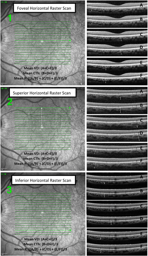
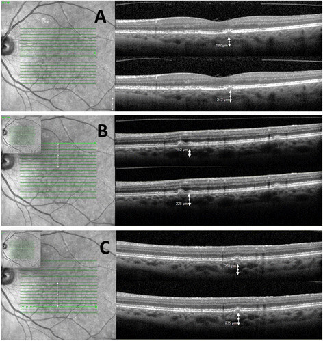
Choroidal thickness (CTh) and choroidal vessel diameter (VD) in the Haler's layer were evaluated as markers of inflammatory insult in non-infectious uveitis (NIU). Spectral-domain optical coherence tomography (Spectralis®, Heidelberg Engineering Inc.) scans were acquired from 23 normal subjects (39 eyes - group 1), 7 subjects with high myopia (14 eyes - group 2), and 19 patients with NIU (23 eyes - group 3). In groups 1 and 2, CTh and VD were measured at 3 different points of the same horizontal OCT scan passing through the fovea and a mean calculated. Mean CTh and VD were calculated in 2 other locations, 2 mm superior and inferior from the chosen foveal horizontal scan. In group 3, three measurements of CTh and VD were obtained within 1 mm of a horizontal scan passing through a retinal lesion; mean CTh and VD were then computed. A ratio (R) between the VD and the corresponding CTh was calculated.

RESULTS

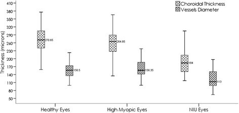
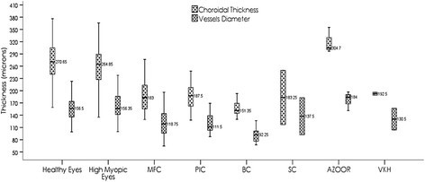
Group 1, 2 and 3 mean age was 29.6, 29.1 and 45.9 years, respectively. Sixteen normal subjects, three myopic subjects and six NIU patients were male.. Group 1 mean CTh did not differ from group 2 (261.6±45.6 vs. 260.2±50.6 µm µm; p>0.05); mean VD was marginally higher in Group 2 (159.8±32.2 vs. 163.2±33.2 µm; p>0.05). Group 3 demonstrated thinner CTh (193.6±54.6 µm) than Groups 1 and 2 (p = 0.02 and <0.001). Group 3 mean VD (123.6±37.4 µm) was also less than that in Groups 1 and 2; the difference was statistically significant only when compared to group 2, p = 0.01. R did not differ across groups (p-values >0.05), indicating that variations in CTh and VD followed the same trend.

CONCLUSIONS

The study reports potential quantitative OCT-derived parameters that may be explored in future trials of non-infectious uveitis. Thinning of choroid and decrease of vessel diameter are observed in patients with chronic NIU compared to controls.

摘要

背景

评估哈勒氏层脉络膜厚度(CTh)和脉络膜血管直径(VD)作为非感染性葡萄膜炎(NIU)炎症损伤的标志物。使用光谱域光学相干断层扫描(Spectralis®,海德堡工程公司)对23名正常受试者(39只眼 - 第1组)、7名高度近视受试者(14只眼 - 第2组)和19名NIU患者(23只眼 - 第3组)进行扫描。在第1组和第2组中,在通过黄斑中心凹的同一水平OCT扫描的3个不同点测量CTh和VD,并计算平均值。在所选黄斑中心凹水平扫描上方和下方2 mm的另外两个位置计算平均CTh和VD。在第3组中,在通过视网膜病变的水平扫描的1 mm范围内获得CTh和VD的三次测量值;然后计算平均CTh和VD。计算VD与相应CTh之间的比值(R)。

结果

第1组、第2组和第3组的平均年龄分别为29.6岁、29.1岁和45.9岁。16名正常受试者、3名近视受试者和6名NIU患者为男性。第1组的平均CTh与第2组无差异(261.6±45.6 vs. 260.2±50.6 µm;p>0.05);第2组的平均VD略高(159.8±32.2 vs. 163.2±33.2 µm;p>0.05)。第3组的CTh(193.6±54.6 µm)比第1组和第2组薄(p = 0.02和<0.001)。第3组的平均VD(123.6±37.4 µm)也低于第1组和第2组;仅与第2组相比差异有统计学意义,p = 0.01。各组之间的R无差异(p值>0.05),表明CTh和VD的变化趋势相同。

结论

该研究报告了潜在的基于OCT的定量参数,可在未来非感染性葡萄膜炎试验中进行探索。与对照组相比,慢性NIU患者观察到脉络膜变薄和血管直径减小。

https://cdn.ncbi.nlm.nih.gov/pmc/blobs/9c89/4884007/f0fc912abe48/12348_2014_Article_14_Fig4_HTML.jpg
https://cdn.ncbi.nlm.nih.gov/pmc/blobs/9c89/4884007/4a898d9673a9/12348_2014_Article_14_Fig1_HTML.jpg
https://cdn.ncbi.nlm.nih.gov/pmc/blobs/9c89/4884007/01b3a3ee45ad/12348_2014_Article_14_Fig2_HTML.jpg
https://cdn.ncbi.nlm.nih.gov/pmc/blobs/9c89/4884007/a3552c47ecbd/12348_2014_Article_14_Fig3_HTML.jpg
https://cdn.ncbi.nlm.nih.gov/pmc/blobs/9c89/4884007/f0fc912abe48/12348_2014_Article_14_Fig4_HTML.jpg

相似文献

1
Variation of choroidal thickness and vessel diameter in patients with posterior non-infectious uveitis.后部非感染性葡萄膜炎患者脉络膜厚度和血管直径的变化
J Ophthalmic Inflamm Infect. 2014 Dec;4(1):14. doi: 10.1186/s12348-014-0014-z. Epub 2014 Aug 16.
2
Full-thickness choroidal thinning as a feature of Fuchs Uveitis Syndrome: quantitative evaluation of the choroid by Enhanced Depth Imaging Optical Coherence Tomography in a cohort of consecutive patients.作为Fuchs葡萄膜炎综合征特征的全层脉络膜变薄:连续患者队列中通过增强深度成像光学相干断层扫描对脉络膜进行定量评估
Graefes Arch Clin Exp Ophthalmol. 2016 Oct;254(10):2025-2031. doi: 10.1007/s00417-016-3475-y. Epub 2016 Sep 2.
3
Choroidal vascular analysis in myopic eyes: evidence of foveal medium vessel layer thinning.近视眼的脉络膜血管分析:黄斑区中血管层变薄的证据。
Int J Retina Vitreous. 2017 May 26;3:28. doi: 10.1186/s40942-017-0081-z. eCollection 2017.
4
Choroidal thickness changes in children with chronic heart failure due to dilated cardiomyopathy.扩张型心肌病所致慢性心力衰竭患儿脉络膜厚度的变化。
Int Ophthalmol. 2021 Jun;41(6):2167-2177. doi: 10.1007/s10792-021-01774-5. Epub 2021 May 9.
5
Evaluation of Retinal and Choroidal Thickness in Fuchs' Uveitis Syndrome.富克斯葡萄膜炎综合征中视网膜和脉络膜厚度的评估
J Ophthalmol. 2016;2016:1657078. doi: 10.1155/2016/1657078. Epub 2016 Aug 8.
6
Retinal and choroidal biometry in highly myopic eyes with spectral-domain optical coherence tomography.高度近视眼视网膜和脉络膜生物测量的光谱域光学相干断层扫描研究
Invest Ophthalmol Vis Sci. 2009 Aug;50(8):3876-80. doi: 10.1167/iovs.08-3325. Epub 2009 Mar 11.
7
The Relationship Between Reticular Macular Disease and Choroidal Thickness.网状黄斑疾病与脉络膜厚度之间的关系。
Curr Eye Res. 2016 Nov;41(11):1492-1497. doi: 10.3109/02713683.2015.1127391. Epub 2016 Apr 26.
8
Comparison of choroidal thicknesses using swept source and spectral domain optical coherence tomography in diseased and normal eyes.使用扫频源光学相干断层扫描和光谱域光学相干断层扫描对患病眼和正常眼的脉络膜厚度进行比较。
Br J Ophthalmol. 2015 Mar;99(3):354-8. doi: 10.1136/bjophthalmol-2014-305331. Epub 2014 Oct 1.
9
QUANTITATIVE ANALYSIS OF STRUCTURAL ALTERATIONS IN THE CHOROID OF PATIENTS WITH ACTIVE BEHÇET UVEITIS.活动性 Behcet 葡萄膜炎患者脉络膜结构改变的定量分析。
Retina. 2018 Apr;38(4):828-840. doi: 10.1097/IAE.0000000000001587.
10
Association Between Parapapillary Choroidal Vessel Density Measured With Optical Coherence Tomography Angiography and Future Visual Field Progression in Patients With Glaucoma.光学相干断层扫描血管造影测量的脉络膜旁血管密度与青光眼患者未来视野进展的相关性。
JAMA Ophthalmol. 2019 Jun 1;137(6):681-688. doi: 10.1001/jamaophthalmol.2019.0422.

引用本文的文献

1
Retinal Structure and Microcirculation Alterations in Patients With Papillary Thyroid Carcinoma: An Optical Coherence Tomography and Angiography Study.甲状腺乳头状癌患者的视网膜结构和微循环改变:一项光学相干断层扫描和血管造影研究
Invest Ophthalmol Vis Sci. 2025 Apr 1;66(4):73. doi: 10.1167/iovs.66.4.73.
2
A Pilot Study Investigating the Relationship Between Choroidal Thickness and Choroidal Vascular Index and Coronary Artery Ectasia.一项关于脉络膜厚度与脉络膜血管指数及冠状动脉扩张之间关系的初步研究。
Diagnostics (Basel). 2025 Jan 26;15(3):286. doi: 10.3390/diagnostics15030286.
3
Effects of inflammation on myopia: evidence and potential mechanisms.

本文引用的文献

1
Choroidal assessment in idiopathic panuveitis using optical coherence tomography.应用光学相干断层扫描评估特发性全葡萄膜炎的脉络膜病变。
Graefes Arch Clin Exp Ophthalmol. 2013 Aug;251(8):2029-36. doi: 10.1007/s00417-013-2330-7. Epub 2013 Mar 28.
2
Characterization of birdshot chorioretinopathy using extramacular enhanced depth optical coherence tomography.应用黄斑外增强深度光学相干断层扫描对鸟枪弹样体视网膜脉络膜病变进行特征描述。
JAMA Ophthalmol. 2013 Mar;131(3):341-50. doi: 10.1001/jamaophthalmol.2013.1724.
3
[Choroidal thickness in normal subjects measured by enhanced depth imaging optical coherence tomography].
炎症对近视的影响:证据与潜在机制。
Front Immunol. 2023 Oct 2;14:1260592. doi: 10.3389/fimmu.2023.1260592. eCollection 2023.
4
Interpreting posterior uveitis by integrating indocyanine green angiography, optical coherence tomography, and optical coherence tomography angiography data: A narrative review.通过整合吲哚菁绿血管造影、光学相干断层扫描和光学相干断层扫描血管造影数据解读后葡萄膜炎:一项叙述性综述
Saudi J Ophthalmol. 2022 Nov 8;36(4):344-355. doi: 10.4103/sjopt.sjopt_69_22. eCollection 2022 Oct-Dec.
5
Efficacy of 0.19 mg Fluocinolone Acetonide Implant in Non-infectious Posterior Uveitis Evaluated as Area Under the Curve.以曲线下面积评估0.19毫克醋酸氟轻松植入剂治疗非感染性后葡萄膜炎的疗效。
Ophthalmol Ther. 2022 Feb;11(1):215-224. doi: 10.1007/s40123-021-00426-2. Epub 2021 Nov 17.
6
Foveal and parafoveal choroidal thickness pattern measuring by swept source optical coherence tomography.应用扫频源光学相干断层扫描测量中心凹及旁中心凹脉络膜厚度。
Eye (Lond). 2019 Sep;33(9):1443-1451. doi: 10.1038/s41433-019-0404-4. Epub 2019 Apr 8.
7
Macular changes in retinal vasculitis.视网膜血管炎中的黄斑改变。
Indian J Ophthalmol. 2018 Mar;66(3):439. doi: 10.4103/ijo.IJO_1232_17.
8
Choroidal thickness in patients with coronary artery disease.冠心病患者的脉络膜厚度
PLoS One. 2017 Jun 20;12(6):e0175691. doi: 10.1371/journal.pone.0175691. eCollection 2017.
9
Choroidal thickness changes determined by EDI-OCT on acute anterior uveitis in patients with HLA-B27-positive ankylosing spondylitis.采用增强深度成像光学相干断层扫描(EDI-OCT)测定HLA-B27阳性强直性脊柱炎患者急性前葡萄膜炎时脉络膜厚度的变化。
Int Ophthalmol. 2018 Feb;38(1):307-312. doi: 10.1007/s10792-017-0464-z. Epub 2017 Feb 14.
10
Choroidal Round Hyporeflectivities in Geographic Atrophy.地图样萎缩中的脉络膜圆形低反射区
PLoS One. 2016 Nov 23;11(11):e0166968. doi: 10.1371/journal.pone.0166968. eCollection 2016.
[通过增强深度成像光学相干断层扫描测量正常受试者的脉络膜厚度]
Zhonghua Yan Ke Za Zhi. 2012 Sep;48(9):819-23.
4
Cellular and physiological mechanisms underlying blood flow regulation in the retina and choroid in health and disease.健康和疾病状态下视网膜和脉络膜血流调节的细胞和生理机制。
Prog Retin Eye Res. 2012 Sep;31(5):377-406. doi: 10.1016/j.preteyeres.2012.04.004. Epub 2012 May 3.
5
Characterization of macular lesions in punctate inner choroidopathy with spectral domain optical coherence tomography.使用频域光学相干断层扫描对点状内层脉络膜病变中的黄斑病变进行特征描述。
J Ophthalmic Inflamm Infect. 2012 Sep;2(3):113-20. doi: 10.1007/s12348-011-0054-6. Epub 2012 Jan 1.
6
Choroidal thickness in healthy Chinese subjects.健康中国受试者的脉络膜厚度。
Invest Ophthalmol Vis Sci. 2011 Dec 20;52(13):9555-60. doi: 10.1167/iovs.11-8076.
7
Subfoveal choroidal thickness in multiple evanescent white dot syndrome.多发性一过性白点综合征的黄斑中心凹下脉络膜厚度
Clin Exp Optom. 2012 Mar;95(2):212-7. doi: 10.1111/j.1444-0938.2011.00668.x. Epub 2011 Oct 24.
8
Enhanced depth imaging optical coherence tomography.增强深度成像光学相干断层扫描
Ophthalmic Surg Lasers Imaging. 2011 Jul;42 Suppl:S75-84. doi: 10.3928/15428877-20110627-07.
9
Macular choroidal thickness and volume in normal subjects measured by swept-source optical coherence tomography.应用扫频源光学相干断层扫描仪测量正常受试者的脉络膜黄斑厚度和体积。
Invest Ophthalmol Vis Sci. 2011 Jul 1;52(8):4971-8. doi: 10.1167/iovs.11-7729.
10
Characteristic spectral-domain optical coherence tomography findings of multifocal choroiditis.多灶性脉络膜炎的特征谱域光相干断层扫描表现。
Retina. 2011 Apr;31(4):717-23. doi: 10.1097/IAE.0b013e318203c1ef.