Verbruggen Stefaan W, Loo Jessica H W, Hayat Tayyib T A, Hajnal Joseph V, Rutherford Mary A, Phillips Andrew T M, Nowlan Niamh C
Department of Bioengineering, Imperial College London, London, SW7 2AZ, UK.
Division of Imaging Sciences, Department of Biomedical Engineering, Kings College London, London, UK.
Biomech Model Mechanobiol. 2016 Aug;15(4):995-1004. doi: 10.1007/s10237-015-0738-1. Epub 2015 Nov 3.
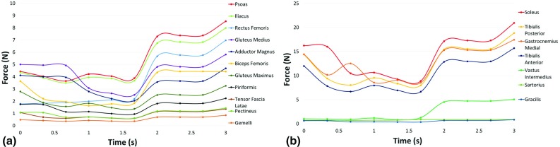
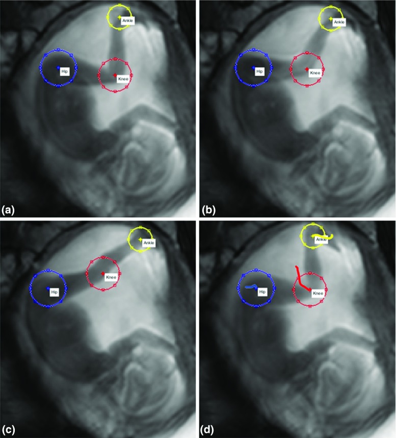
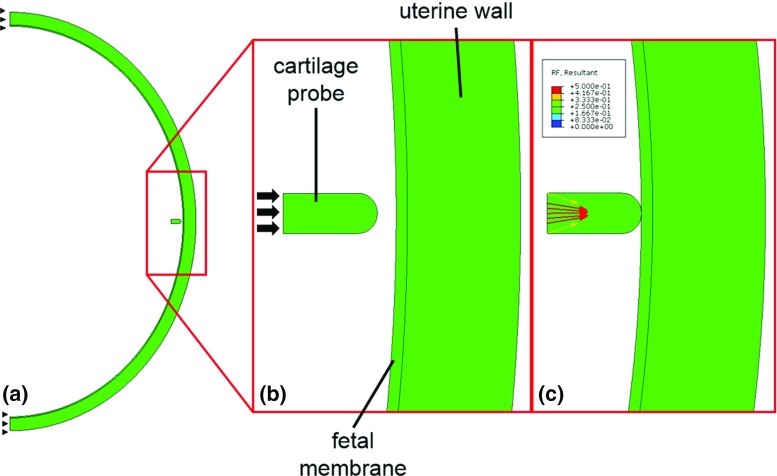
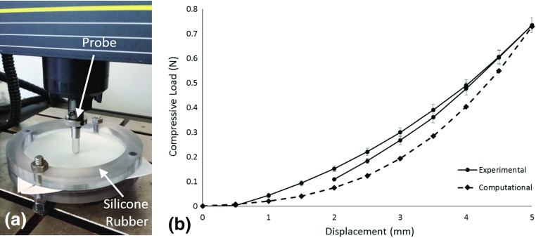
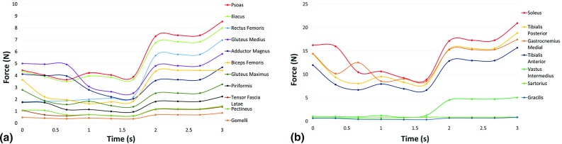
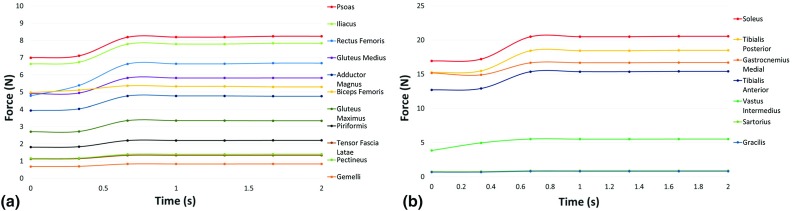
Fetal movements in the uterus are a natural part of development and are known to play an important role in normal musculoskeletal development. However, very little is known about the biomechanical stimuli that arise during movements in utero, despite these stimuli being crucial to normal bone and joint formation. Therefore, the objective of this study was to create a series of computational steps by which the forces generated during a kick in utero could be predicted from clinically observed fetal movements using novel cine-MRI data of three fetuses, aged 20-22 weeks. A custom tracking software was designed to characterize the movements of joints in utero, and average uterus deflection of [Formula: see text] mm due to kicking was calculated. These observed displacements provided boundary conditions for a finite element model of the uterine environment, predicting an average reaction force of [Formula: see text] N generated by a kick against the uterine wall. Finally, these data were applied as inputs for a musculoskeletal model of a fetal kick, resulting in predicted maximum forces in the muscles surrounding the hip joint of approximately 8 N, while higher maximum forces of approximately 21 N were predicted for the muscles surrounding the knee joint. This study provides a novel insight into the closed mechanical environment of the uterus, with an innovative method allowing elucidation of the biomechanical interaction of the developing fetus with its surroundings.
子宫内的胎儿运动是发育过程中的自然组成部分,已知其在正常肌肉骨骼发育中发挥重要作用。然而,尽管这些刺激对正常骨骼和关节形成至关重要,但对于子宫内运动期间产生的生物力学刺激却知之甚少。因此,本研究的目的是创建一系列计算步骤,通过使用三个年龄在20 - 22周的胎儿的新型电影磁共振成像(cine - MRI)数据,从临床观察到的胎儿运动中预测子宫内踢腿时产生的力。设计了一个定制的跟踪软件来表征子宫内关节的运动,并计算出由于踢腿导致的平均子宫偏转[公式:见原文]毫米。这些观察到的位移为子宫环境的有限元模型提供了边界条件,预测出一次踢腿对子宫壁产生的平均反作用力为[公式:见原文]牛顿。最后,将这些数据用作胎儿踢腿肌肉骨骼模型的输入,得出髋关节周围肌肉的预测最大力约为8牛顿,而膝关节周围肌肉的预测最大力更高,约为21牛顿。本研究为子宫的封闭力学环境提供了新的见解,采用了一种创新方法来阐明发育中的胎儿与其周围环境的生物力学相互作用。