Stetson Powell Orthopaedics and Sports Medicine, Burbank, California, USA.
Orthop J Sports Med. 2014 Jul 2;2(7):2325967114540407. doi: 10.1177/2325967114540407. eCollection 2014 Jul.
Many studies have compared the diagnostic capabilities of low-field magnetic resonance imaging (MRI) scanners to high-field MRI scanners; however, few have evaluated the low-field MRI diagnoses compared with intraoperative findings.
To determine the accuracy and sensitivity of low-field MRI scanners in diagnosing lesions of the rotator cuff and glenoid labrum.
Cohort study (diagnosis); Level of evidence, 3.
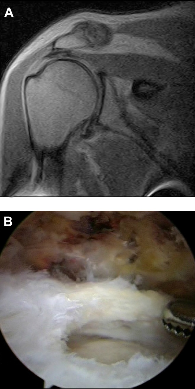
Over a 2-year period, MRI examinations without intra-articular contrast were performed on 79 patients for shoulder pathologies using an in-office 0.2-T extremity scanner. The MRI examinations were read by board-certified, musculoskeletal fellowship-trained radiologists. All patients underwent shoulder arthroscopy performed by a single sports fellowship-trained orthopaedic surgeon within a mean time of 56 days (range, 8-188 days) after the MRI examination. The mean patient age was 54 years (range, 18-81 years). Operative notes from the shoulder arthroscopies were then retrospectively reviewed by a single blinded observer, and the intraoperative findings were compared with the MRI reports.
For partial-thickness rotator cuff tears, the sensitivity, specificity, positive predictive value, and negative predictive value were 85%, 89%, 79%, and 92%, respectively. For full-thickness rotator cuff tears, the respective values were 97%, 100%, 100%, and 98%. For anterior labral lesions, the values were 86%, 99%, 86%, and 99%, and for superior labral anterior-posterior (SLAP) lesions, the values were 20%, 100%, 100%, and 79%, respectively.
Low-field MRI is an accurate tool for evaluation of partial- and full-thickness rotator cuff tears; however, it is not effective in diagnosing SLAP lesions. More information is needed to properly assess its ability to diagnose anterior and posterior labral lesions.
许多研究比较了低磁场磁共振成像(MRI)扫描仪与高磁场 MRI 扫描仪的诊断能力;然而,很少有研究评估低磁场 MRI 诊断与术中发现的比较。
确定低磁场 MRI 扫描仪在诊断肩袖和盂唇病变中的准确性和敏感性。
队列研究(诊断);证据水平,3 级。
在 2 年期间,使用办公室内的 0.2-T 肢体扫描仪对 79 例肩部病变患者进行了无关节内对比的 MRI 检查。MRI 检查由经过董事会认证、具有肌肉骨骼专业 fellowship培训的放射科医生进行阅读。所有患者均在 MRI 检查后平均 56 天(范围为 8-188 天)内由一位具有运动专业 fellowship培训的骨科医生进行了肩关节镜检查。患者平均年龄为 54 岁(范围为 18-81 岁)。然后,由一位单独的盲法观察者回顾性审查肩关节镜手术记录,并将术中发现与 MRI 报告进行比较。
对于部分厚度肩袖撕裂,敏感性、特异性、阳性预测值和阴性预测值分别为 85%、89%、79%和 92%。对于全层肩袖撕裂,相应的值分别为 97%、100%、100%和 98%。对于前盂唇病变,其值分别为 86%、99%、86%和 99%,而对于上盂唇前-后(SLAP)病变,其值分别为 20%、100%、100%和 79%。
低磁场 MRI 是评估部分和全层肩袖撕裂的准确工具;然而,它在诊断 SLAP 病变方面效果不佳。需要更多信息来正确评估其诊断前、后盂唇病变的能力。