Stornetta Ruth L, Inglis M Andrews, Viar Kenneth E, Guyenet Patrice G
Department of Pharmacology, University of Virginia Health System, 1340 Jefferson Park Avenue, P.O. Box 800735, Charlottesville, VA, 22908-0735, USA.
Brain Struct Funct. 2016 Nov;221(8):4027-4044. doi: 10.1007/s00429-015-1143-3. Epub 2015 Nov 11.
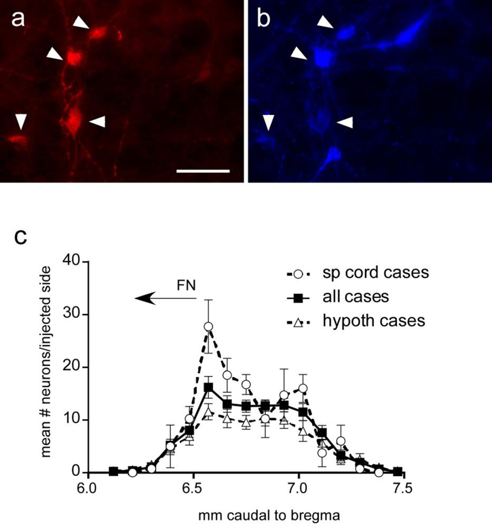
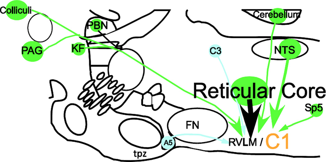
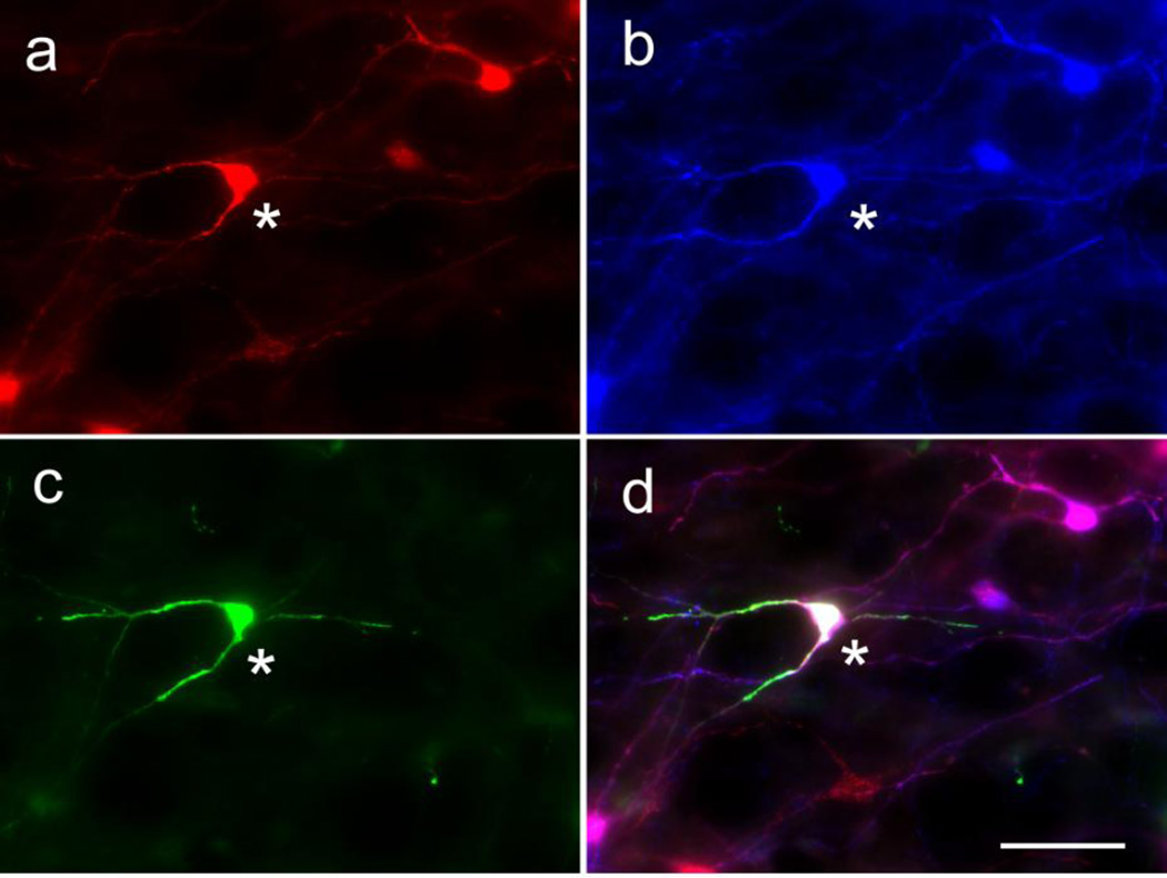
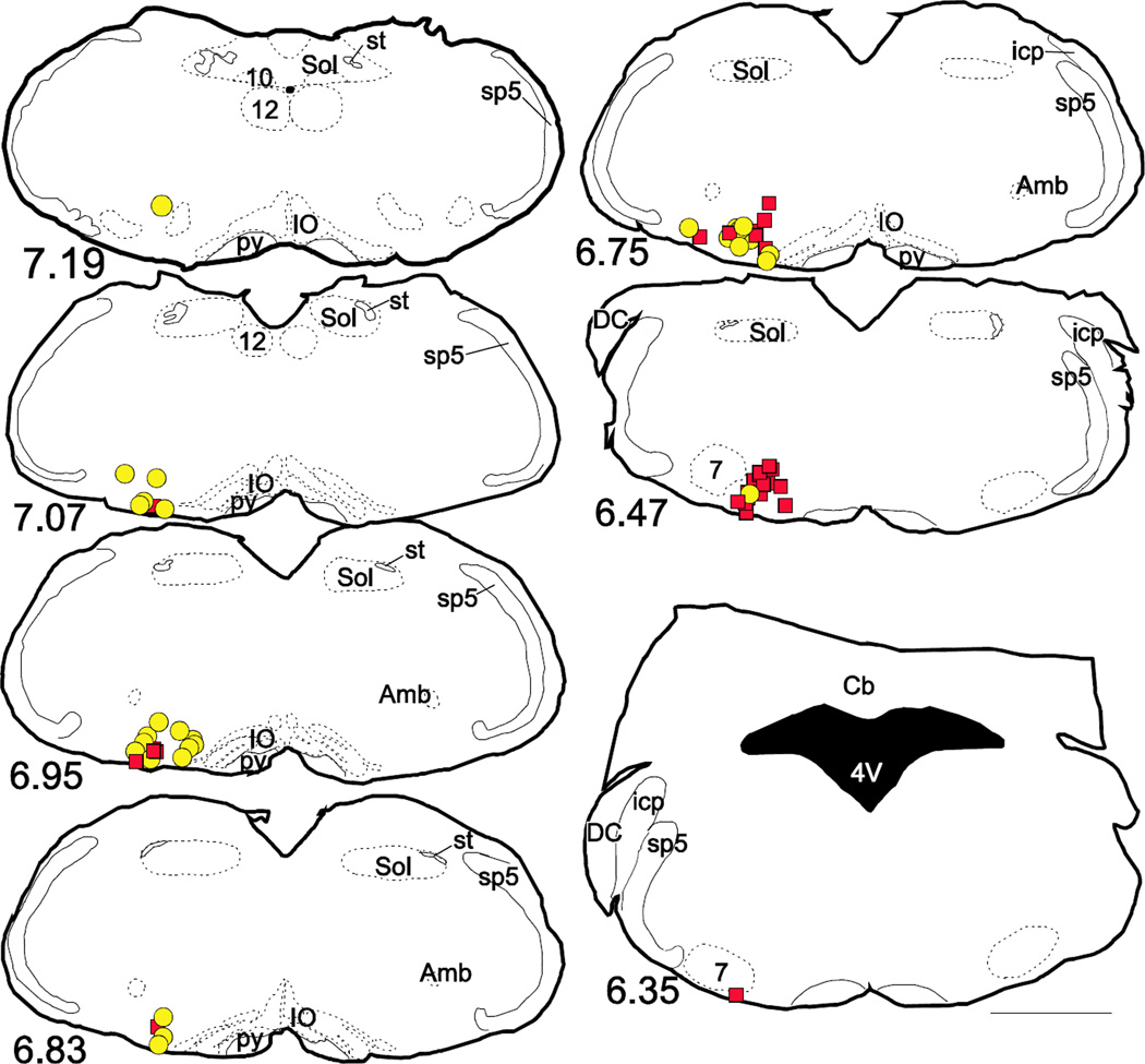
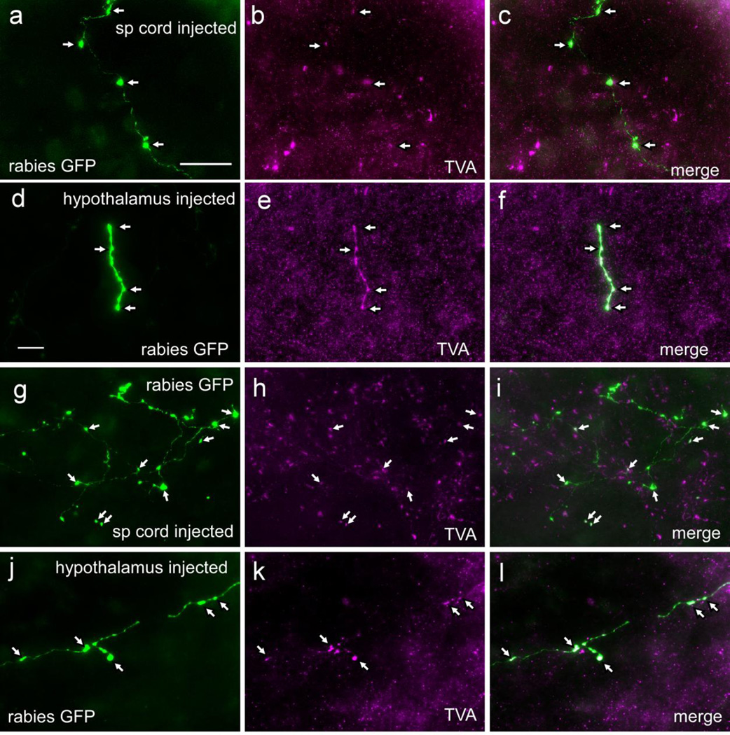
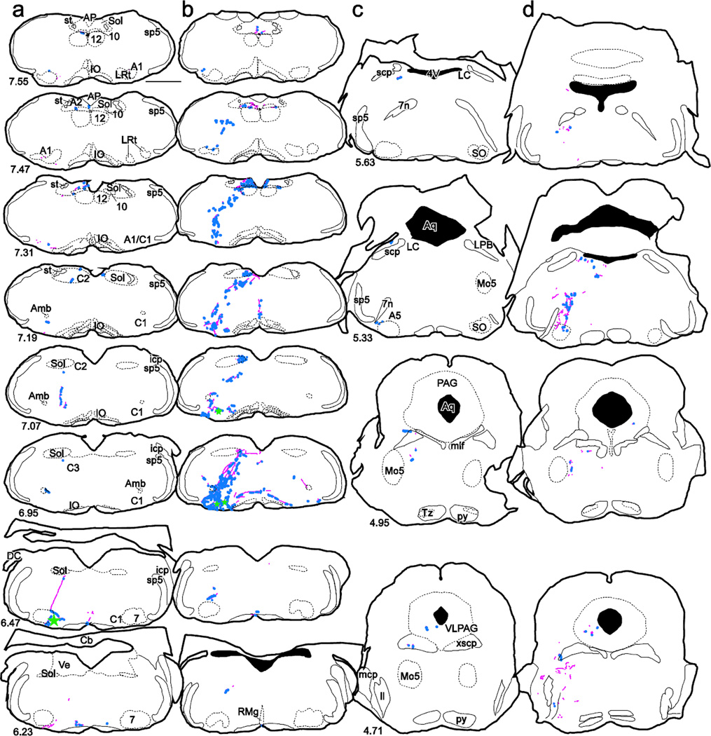
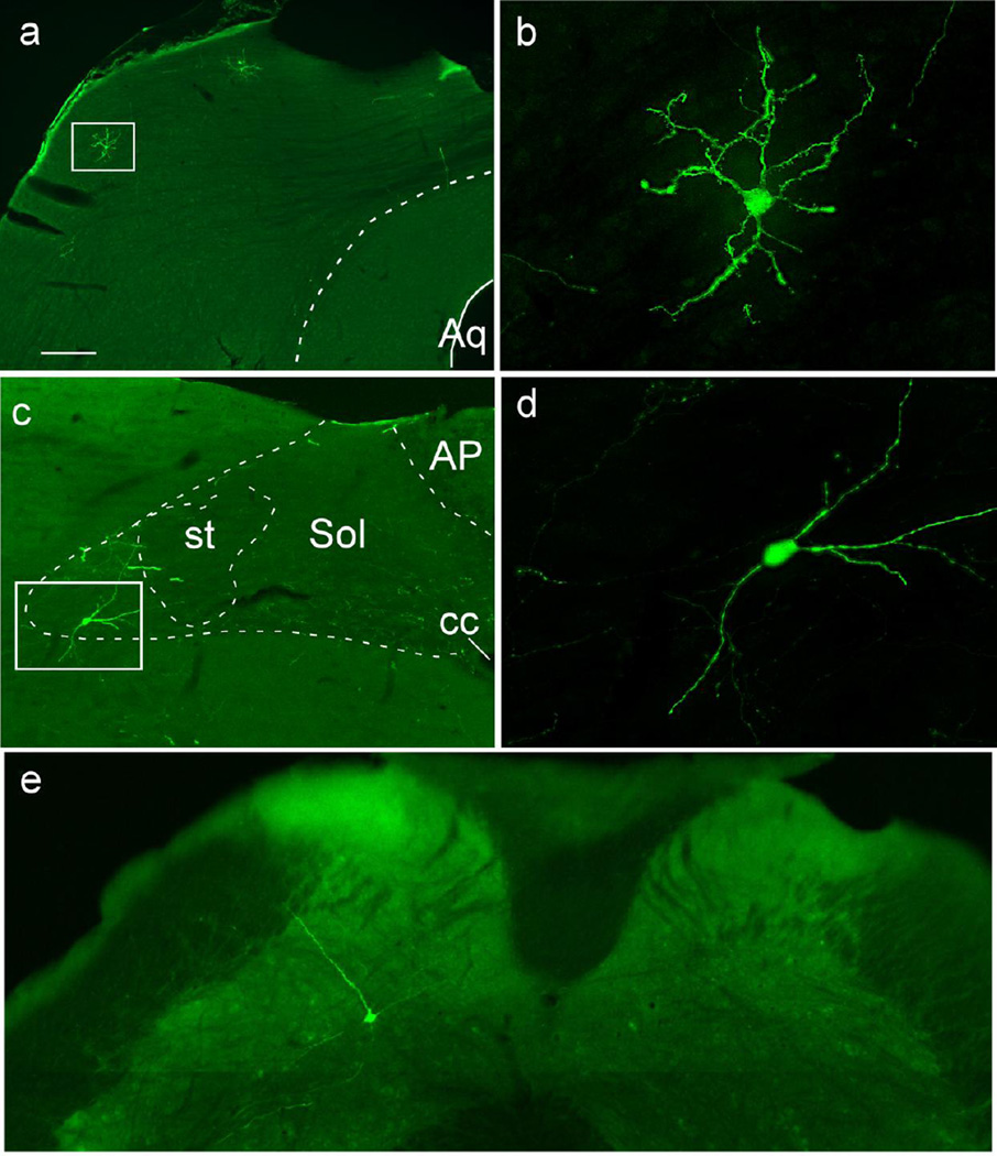
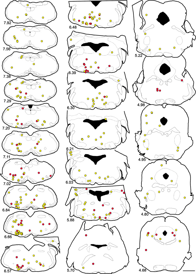
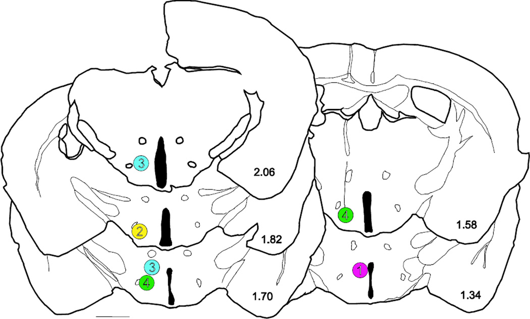
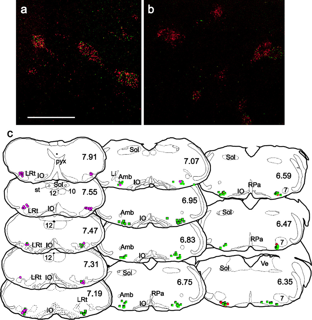
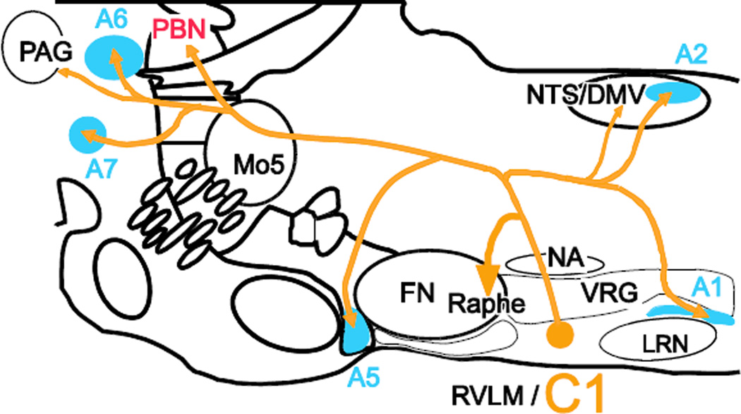
The axonal projections and synaptic input of the C1 adrenergic neurons of the rostral ventrolateral medulla (VLM) were examined using transgenic dopamine-beta hydroxylase Cre mice and modified rabies virus. Cre-dependent viral vectors expressing TVA (receptor for envelopeA) and rabies glycoprotein were injected into the left VLM. EnvelopeA-pseudotyped rabies-EGFP glycoprotein-deficient virus (rabies-EGFP) was injected 4-6 weeks later in either thoracic spinal cord (SC) or hypothalamus. TVA immunoreactivity was detected almost exclusively (95 %) in VLM C1 neurons. In mice with SC injections of rabies-EGFP, starter cells (expressing TVA + EGFP) were found at the rostral end of the VLM; in mice with hypothalamic injections starter C1 cells were located more caudally. C1 neurons innervating SC or hypothalamus had other terminal fields in common (e.g., dorsal vagal complex, locus coeruleus, raphe pallidus and periaqueductal gray matter). Putative inputs to C1 cells with SC or hypothalamic projections originated from the same brain regions, especially the lower brainstem reticular core from spinomedullary border to rostral pons. Putative input neurons to C1 cells were also observed in the nucleus of the solitary tract, caudal VLM, caudal spinal trigeminal nucleus, cerebellum, periaqueductal gray matter and inferior and superior colliculi. In sum, regardless of whether they innervate SC or hypothalamus, VLM C1 neurons receive input from the same general brain regions. One interpretation is that many types of somatic or internal stimuli recruit these neurons en bloc to produce a stereotyped acute stress response with sympathetic, parasympathetic, vigilance and neuroendocrine components.
利用转基因多巴胺-β羟化酶Cre小鼠和改良狂犬病毒,研究了延髓头端腹外侧区(VLM)C1肾上腺素能神经元的轴突投射和突触输入。将表达TVA(包膜A受体)和狂犬病毒糖蛋白的Cre依赖性病毒载体注入左侧VLM。4至6周后,将包膜A假型化的狂犬病毒增强绿色荧光蛋白糖蛋白缺陷病毒(狂犬病毒-EGFP)注入胸段脊髓(SC)或下丘脑。TVA免疫反应几乎仅在VLM C1神经元中检测到(95%)。在向SC注射狂犬病毒-EGFP的小鼠中,起始细胞(表达TVA + EGFP)位于VLM的头端;在下丘脑注射的小鼠中,起始C1细胞位于更靠尾侧的位置。支配SC或下丘脑的C1神经元有其他共同的终末区域(如迷走背核、蓝斑、中缝苍白核和导水管周围灰质)。投射到SC或下丘脑的C1细胞的假定输入起源于相同的脑区,特别是从脊髓延髓边界到脑桥头端的低位脑干网状核心。在孤束核、尾侧VLM、尾侧三叉神经脊束核、小脑、导水管周围灰质以及上、下丘中也观察到了C1细胞的假定输入神经元。总之,无论VLM C1神经元支配SC还是下丘脑,它们都从相同的一般脑区接收输入。一种解释是,许多类型的躯体或内部刺激会整体募集这些神经元,以产生一种具有交感、副交感、警觉和神经内分泌成分的定型急性应激反应。