Chang Feng-Yu, Tsai Meng-Tsan, Wang Zu-Yi, Chi Chun-Kai, Lee Cheng-Kuang, Yang Chih-Hsun, Chan Ming-Che, Lee Ya-Ju
Department of Electrical Engineering, Chang Gung University, 259, Wen-Hwa 1st Rd., Kwei-Shan Dist., Taoyuan city, 33302, Taiwan.
Medical Imaging Research Center, Institute for Radiological Research, Chang Gung University and Chang Gung Memorial Hospital at Linkou, Taoyuan, Taiwan.
Sci Rep. 2015 Nov 16;5:16739. doi: 10.1038/srep16739.
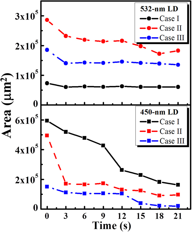
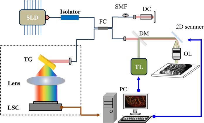
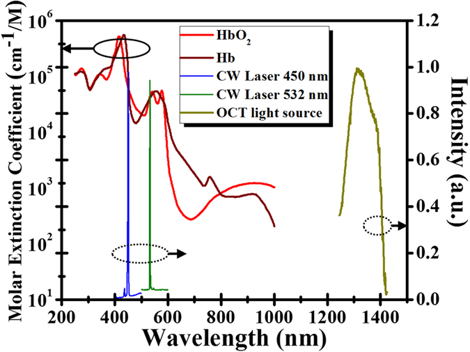
Blood coagulation is the clotting and subsequent dissolution of the clot following repair to the damaged tissue. However, inducing blood coagulation is difficult for some patients with homeostasis dysfunction or during surgery. In this study, we proposed a method to develop an integrated system that combines optical coherence tomography (OCT) and laser microsurgery for blood coagulation. Also, an algorithm for positioning of the treatment location from OCT images was developed. With OCT scanning, 2D/3D OCT images and angiography of tissue can be obtained simultaneously, enabling to noninvasively reconstruct the morphological and microvascular structures for real-time monitoring of changes in biological tissues during laser microsurgery. Instead of high-cost pulsed lasers, continuous-wave laser diodes (CW-LDs) with the central wavelengths of 450 nm and 532 nm are used for blood coagulation, corresponding to higher absorption coefficients of oxyhemoglobin and deoxyhemoglobin. Experimental results showed that the location of laser exposure can be accurately controlled with the proposed approach of imaging-based feedback positioning. Moreover, blood coagulation can be efficiently induced by CW-LDs and the coagulation process can be monitored in real-time with OCT. This technology enables to potentially provide accurate positioning for laser microsurgery and control the laser exposure to avoid extra damage by real-time OCT imaging.
血液凝固是指在受损组织修复后,血液形成凝块并随后溶解凝块的过程。然而,对于一些内稳态功能失调的患者或在手术过程中,诱导血液凝固是困难的。在本研究中,我们提出了一种开发集成系统的方法,该系统将光学相干断层扫描(OCT)和激光显微手术相结合用于血液凝固。此外,还开发了一种从OCT图像定位治疗位置的算法。通过OCT扫描,可以同时获得组织的二维/三维OCT图像和血管造影,从而能够无创地重建形态和微血管结构,以实时监测激光显微手术期间生物组织的变化。代替高成本的脉冲激光器,使用中心波长为450 nm和532 nm的连续波激光二极管(CW-LD)进行血液凝固,这对应于氧合血红蛋白和脱氧血红蛋白的较高吸收系数。实验结果表明,采用所提出的基于成像的反馈定位方法可以精确控制激光照射的位置。此外,CW-LD可以有效地诱导血液凝固,并且可以用OCT实时监测凝固过程。该技术有可能为激光显微手术提供精确的定位,并通过实时OCT成像控制激光照射以避免额外损伤。