• 文献检索
  • 文档翻译
  • 深度研究
  • 学术资讯
  • Suppr Zotero 插件Zotero 插件
  • 邀请有礼
  • 套餐&价格
  • 历史记录
应用&插件
Suppr Zotero 插件Zotero 插件浏览器插件Mac 客户端Windows 客户端微信小程序
定价
高级版会员购买积分包购买API积分包
服务
文献检索文档翻译深度研究API 文档MCP 服务
关于我们
关于 Suppr公司介绍联系我们用户协议隐私条款
关注我们

Suppr 超能文献

核心技术专利:CN118964589B侵权必究
粤ICP备2023148730 号-1Suppr @ 2026

文献检索

告别复杂PubMed语法,用中文像聊天一样搜索,搜遍4000万医学文献。AI智能推荐,让科研检索更轻松。

立即免费搜索

文件翻译

保留排版,准确专业,支持PDF/Word/PPT等文件格式,支持 12+语言互译。

免费翻译文档

深度研究

AI帮你快速写综述,25分钟生成高质量综述,智能提取关键信息,辅助科研写作。

立即免费体验

光学相干断层扫描引导下的异种移植模型中的激光脑癌手术。

Laser brain cancer surgery in a xenograft model guided by optical coherence tomography.

机构信息

University of Texas at Austin.

University of Texas Health Science Center at San Antonio.

出版信息

Theranostics. 2019 May 26;9(12):3555-3564. doi: 10.7150/thno.31811. eCollection 2019.

DOI:10.7150/thno.31811
PMID:31281497
原文链接:https://pmc.ncbi.nlm.nih.gov/articles/PMC6587169/
Abstract

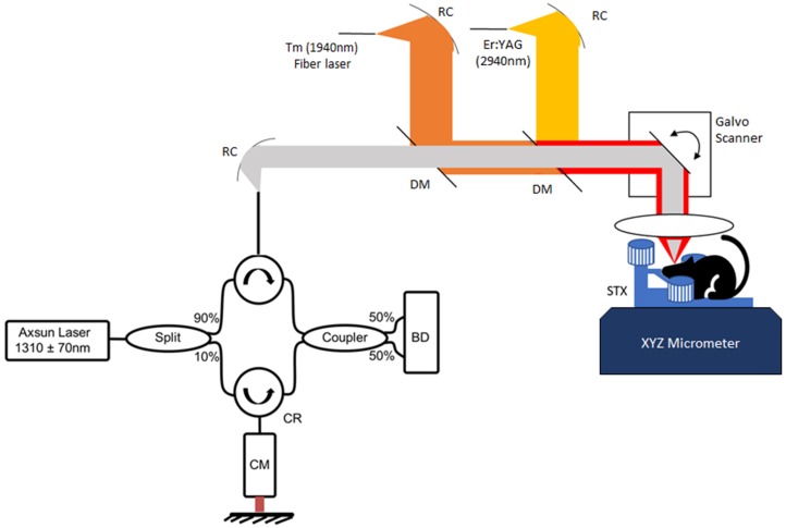
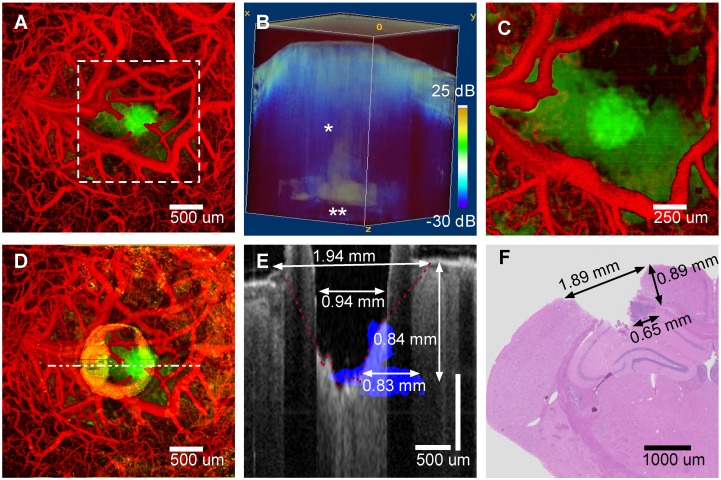
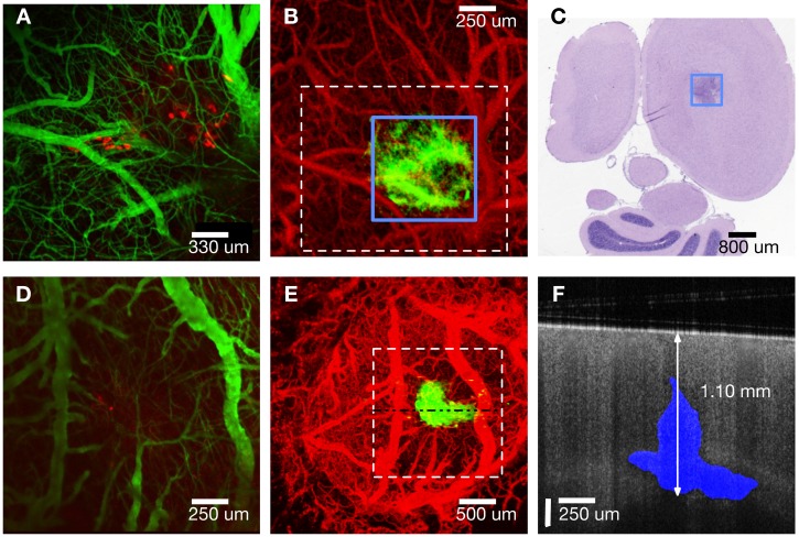
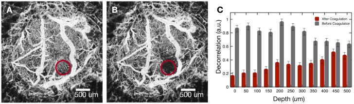
Higher precision surgical devices are needed for tumor resections near critical brain structures. The goal of this study is to demonstrate feasibility of a system capable of precise and bloodless tumor ablation. An image-guided laser surgical system is presented for excision of brain tumors in a murine xenograft model. The system combines optical coherence tomography (OCT) guidance with surgical lasers for high-precision tumor ablation (Er:YAG) and microcirculation coagulation (Thulium (Tm) fiber laser). A fluorescent human glioblastoma cell line was injected into mice and allowed to grow four weeks. Craniotomies were performed and tumors were imaged with confocal fluorescence microscopy. The mice were subsequently OCT imaged prior, during and after laser coagulation and/or ablation. The prior OCT images were used to compute three-dimensional tumor margin and angiography images, which guided the coagulation and ablation steps. Histology of the treated regions was then compared to post-treatment OCT images. Tumor sizing based on OCT margin detection matched histology to within experimental error. Although fluorescence microscopy imaging showed the tumors were collocated with OCT imaging, margin assessment using confocal microscopy failed to see the extent of the tumor beyond ~ 250 µm in depth, as verified by OCT and histology. The two-laser approach to surgery utilizing Tm wavelength for coagulation and Er:YAG for ablation yielded bloodless resection of tumor regions with minimal residual damage as seen in histology. Precise and bloodless tumor resection under OCT image guidance is demonstrated in the murine xenograft brain cancer model. Tumor margins and vasculature are accurately made visible without need for exogenous contrast agents.

摘要

需要更高精度的手术器械来进行靠近关键脑结构的肿瘤切除。本研究的目的是展示一种能够精确进行无血肿瘤消融的系统的可行性。提出了一种用于在鼠异种移植模型中切除脑肿瘤的图像引导激光手术系统。该系统将光学相干断层扫描(OCT)引导与手术激光结合,用于高精度肿瘤消融(Er:YAG)和微循环凝固(Tm 光纤激光)。将荧光人类胶质母细胞瘤细胞系注射到小鼠体内并使其生长四周。进行开颅术,并使用共聚焦荧光显微镜对肿瘤进行成像。随后对小鼠进行 OCT 成像,在激光凝固和/或消融之前、期间和之后进行。之前的 OCT 图像用于计算三维肿瘤边缘和血管造影图像,这些图像指导了凝固和消融步骤。然后将处理区域的组织学与治疗后的 OCT 图像进行比较。基于 OCT 边缘检测的肿瘤大小与组织学匹配,误差在实验范围内。尽管荧光显微镜成像显示肿瘤与 OCT 成像共定位,但共聚焦显微镜的边缘评估未能看到肿瘤在深度超过约 250 µm 的范围,这通过 OCT 和组织学得到了验证。利用 Tm 波长进行凝固和 Er:YAG 进行消融的双激光手术方法实现了肿瘤区域的无血切除,组织学上可见最小的残留损伤。在鼠异种移植脑癌模型中证明了在 OCT 图像引导下进行精确、无血的肿瘤切除。肿瘤边缘和脉管系统无需外源性对比剂即可准确显现。

https://cdn.ncbi.nlm.nih.gov/pmc/blobs/50ee/6587169/b0538a63640f/thnov09p3555g004.jpg
https://cdn.ncbi.nlm.nih.gov/pmc/blobs/50ee/6587169/34f7afa621eb/thnov09p3555g001.jpg
https://cdn.ncbi.nlm.nih.gov/pmc/blobs/50ee/6587169/2a3229c677c8/thnov09p3555g002.jpg
https://cdn.ncbi.nlm.nih.gov/pmc/blobs/50ee/6587169/55c5c10657bb/thnov09p3555g003.jpg
https://cdn.ncbi.nlm.nih.gov/pmc/blobs/50ee/6587169/b0538a63640f/thnov09p3555g004.jpg

相似文献

1
Laser brain cancer surgery in a xenograft model guided by optical coherence tomography.光学相干断层扫描引导下的异种移植模型中的激光脑癌手术。
Theranostics. 2019 May 26;9(12):3555-3564. doi: 10.7150/thno.31811. eCollection 2019.
2
Er:YAG laser brain surgery with vascular specific coagulation.铒:YAG 激光脑外科手术伴血管特异性凝固。
Lasers Surg Med. 2022 Oct;54(8):1107-1115. doi: 10.1002/lsm.23591. Epub 2022 Aug 10.
3
Bioluminescence imaging and two-photon microscopy guided laser ablation of GBM decreases tumor burden.生物发光成像和双光子显微镜引导下的 GBM 激光消融可降低肿瘤负担。
Theranostics. 2018 Jul 16;8(15):4072-4085. doi: 10.7150/thno.25357. eCollection 2018.
4
A novel integration of spectral-domain optical-coherence-tomography and laser-ablation system for precision treatment.一种新型的光谱域光学相干层析成像和激光烧蚀系统的集成,用于精确治疗。
Int J Comput Assist Radiol Surg. 2018 Mar;13(3):411-423. doi: 10.1007/s11548-017-1664-8. Epub 2017 Sep 9.
5
Intelligent optical diagnosis and treatment system for automated image-guided laser ablation of tumors.肿瘤自动图像引导激光消融的智能光学诊断和治疗系统。
Int J Comput Assist Radiol Surg. 2021 Dec;16(12):2147-2157. doi: 10.1007/s11548-021-02457-3. Epub 2021 Aug 7.
6
Optical coherence tomography image-guided smart laser knife for surgery.用于手术的光学相干断层扫描图像引导智能激光刀
Lasers Surg Med. 2018 Mar;50(3):202-212. doi: 10.1002/lsm.22705. Epub 2017 Aug 7.
7
Fiber-laser platform for precision brain surgery.用于精准脑部手术的光纤激光平台。
Biomed Opt Express. 2022 Mar 9;13(4):1985-1994. doi: 10.1364/BOE.449312. eCollection 2022 Apr 1.
8
A Fluorescence-Guided Laser Ablation System for Removal of Residual Cancer in a Mouse Model of Soft Tissue Sarcoma.一种用于软组织肉瘤小鼠模型中去除残留癌组织的荧光引导激光消融系统。
Theranostics. 2016 Jan 1;6(2):155-66. doi: 10.7150/thno.13536. eCollection 2016.
9
Imaging of human brain tumor tissue by near-infrared laser coherence tomography.近红外激光相干断层扫描对人脑肿瘤组织的成像
Acta Neurochir (Wien). 2009 May;151(5):507-17; discussion 517. doi: 10.1007/s00701-009-0248-y. Epub 2009 Apr 3.
10
High-resolution optical coherence tomography-guided laser ablation of surgical tissue.高分辨率光学相干断层扫描引导下的手术组织激光消融
J Surg Res. 1999 Apr;82(2):275-84. doi: 10.1006/jsre.1998.5555.

引用本文的文献

1
Research progress on the use of the optical coherence tomography system for the diagnosis and treatment of central nervous system tumors.光学相干断层扫描系统在中枢神经系统肿瘤诊断与治疗中的应用研究进展
Ibrain. 2024 Nov 22;11(1):3-18. doi: 10.1002/ibra.12184. eCollection 2025 Spring.
2
Optical coherence tomography (OCT) and OCT angiography: Technological development and applications in brain science.光学相干断层扫描(OCT)与OCT血管造影:技术发展及其在脑科学中的应用
Theranostics. 2025 Jan 1;15(1):122-140. doi: 10.7150/thno.97192. eCollection 2025.
3
The LMIT: Light-mediated minimally-invasive theranostics in oncology.

本文引用的文献

1
Quantitative nontumorous and tumorous human brain tissue assessment using microstructural co- and cross-polarized optical coherence tomography.利用微观结构共极化和交叉极化光相干断层扫描技术对非肿瘤和肿瘤性人脑组织进行定量评估。
Sci Rep. 2019 Feb 14;9(1):2024. doi: 10.1038/s41598-019-38493-y.
2
An Intelligent Theranostics Method Using Optical Coherence Tomography Guided Automatic Laser Ablation for Neurosurgery.一种使用光学相干断层扫描引导自动激光消融的智能诊疗方法用于神经外科手术。
Annu Int Conf IEEE Eng Med Biol Soc. 2018 Jul;2018:3224-3227. doi: 10.1109/EMBC.2018.8513016.
3
Real-time visualization and interaction with static and live optical coherence tomography volumes in immersive virtual reality.
LMIT:肿瘤光介导的微创治疗与诊断。
Theranostics. 2024 Jan 1;14(1):341-362. doi: 10.7150/thno.87783. eCollection 2024.
4
Polarization-sensitive optical coherence tomography for renal tumor detection in human kidneys.用于检测人肾中肾肿瘤的偏振敏感光学相干断层扫描技术。
Opt Lasers Eng. 2024 Feb;173. doi: 10.1016/j.optlaseng.2023.107900. Epub 2023 Oct 23.
5
Real-time closed-loop tissue-specific laser osteotomy using deep-learning-assisted optical coherence tomography.使用深度学习辅助光学相干断层扫描的实时闭环组织特异性激光截骨术。
Biomed Opt Express. 2023 May 31;14(6):2986-3002. doi: 10.1364/BOE.486660. eCollection 2023 Jun 1.
6
Pulsed thulium laser blood vessel haemostasis as an alternative to bipolar forceps during neurosurgical tumour resection.经皮脉冲铥激光血管止血术作为神经外科肿瘤切除中双极电凝镊的替代方法。
Lasers Med Sci. 2023 Mar 27;38(1):94. doi: 10.1007/s10103-023-03747-9.
7
Towards OCT-Guided Endoscopic Laser Surgery-A Review.迈向光学相干断层扫描引导的内镜激光手术——综述
Diagnostics (Basel). 2023 Feb 11;13(4):677. doi: 10.3390/diagnostics13040677.
8
Safeness and efficacy of 2-µm handheld thulium laser during microsurgical resection of supratentorial and infratentorial meningiomas: Experience of a single center.2微米手持式铥激光在幕上和幕下脑膜瘤显微手术切除中的安全性和有效性:单中心经验
Front Surg. 2022 Dec 16;9:1021019. doi: 10.3389/fsurg.2022.1021019. eCollection 2022.
9
Research progress on the application of optical coherence tomography in the field of oncology.光学相干断层扫描在肿瘤学领域应用的研究进展
Front Oncol. 2022 Jul 25;12:953934. doi: 10.3389/fonc.2022.953934. eCollection 2022.
10
Laser coagulation and hemostasis of large diameter blood vessels: effect of shear stress and flow velocity.激光凝固和大直径血管止血:剪切应力和流速的影响。
Sci Rep. 2022 May 19;12(1):8375. doi: 10.1038/s41598-022-12128-1.
在沉浸式虚拟现实中对静态和动态光学相干断层扫描容积进行实时可视化和交互。
Biomed Opt Express. 2018 May 30;9(6):2825-2843. doi: 10.1364/BOE.9.002825. eCollection 2018 Jun 1.
4
Bioluminescence imaging and two-photon microscopy guided laser ablation of GBM decreases tumor burden.生物发光成像和双光子显微镜引导下的 GBM 激光消融可降低肿瘤负担。
Theranostics. 2018 Jul 16;8(15):4072-4085. doi: 10.7150/thno.25357. eCollection 2018.
5
Automated differentiation between meningioma and healthy brain tissue based on optical coherence tomography ex vivo images using texture features.基于纹理特征的光学相干断层扫描离体图像对脑膜瘤与正常脑组织的自动区分。
J Biomed Opt. 2018 Feb;23(7):1-7. doi: 10.1117/1.JBO.23.7.071205.
6
Optical coherence tomography for precision brain imaging, neurosurgical guidance and minimally invasive theranostics.光学相干断层扫描在精准脑部成像、神经外科指导和微创治疗中的应用。
Biosci Trends. 2018 Mar 18;12(1):12-23. doi: 10.5582/bst.2017.01258. Epub 2018 Jan 15.
7
A novel integration of spectral-domain optical-coherence-tomography and laser-ablation system for precision treatment.一种新型的光谱域光学相干层析成像和激光烧蚀系统的集成,用于精确治疗。
Int J Comput Assist Radiol Surg. 2018 Mar;13(3):411-423. doi: 10.1007/s11548-017-1664-8. Epub 2017 Sep 9.
8
Optical coherence tomography image-guided smart laser knife for surgery.用于手术的光学相干断层扫描图像引导智能激光刀
Lasers Surg Med. 2018 Mar;50(3):202-212. doi: 10.1002/lsm.22705. Epub 2017 Aug 7.
9
Robust and fast characterization of OCT-based optical attenuation using a novel frequency-domain algorithm for brain cancer detection.使用基于 OCT 的新型频域算法对脑癌进行快速、稳健的光学衰减特征分析。
Sci Rep. 2017 Mar 22;7:44909. doi: 10.1038/srep44909.
10
Optical coherence tomography based angiography [Invited].基于光学相干断层扫描的血管造影术[特邀报告]
Biomed Opt Express. 2017 Jan 24;8(2):1056-1082. doi: 10.1364/BOE.8.001056. eCollection 2017 Feb 1.