Katorcha Elizaveta, Klimova Nina, Makarava Natallia, Savtchenko Regina, Pan Xuefang, Annunziata Ida, Takahashi Kohta, Miyagi Taeko, Pshezhetsky Alexey V, d'Azzo Alessandra, Baskakov Ilia V
Center for Biomedical Engineering and Technology, University of Maryland School of Medicine, Baltimore, Maryland, United States of America.
Department of Anatomy and Neurobiology, University of Maryland School of Medicine, Baltimore, Maryland, United States of America.
PLoS One. 2015 Nov 16;10(11):e0143218. doi: 10.1371/journal.pone.0143218. eCollection 2015.
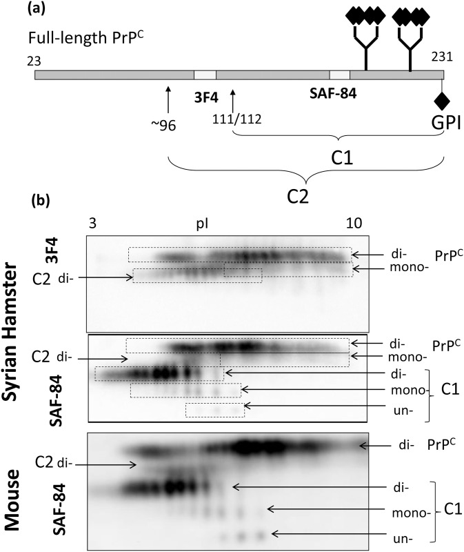
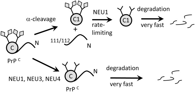
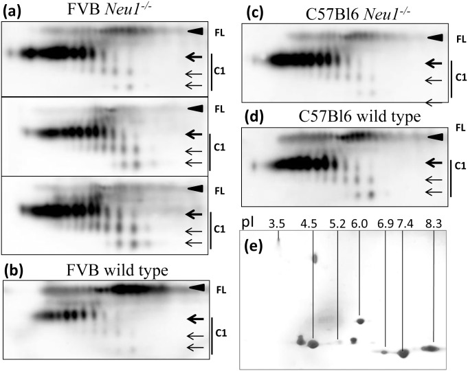
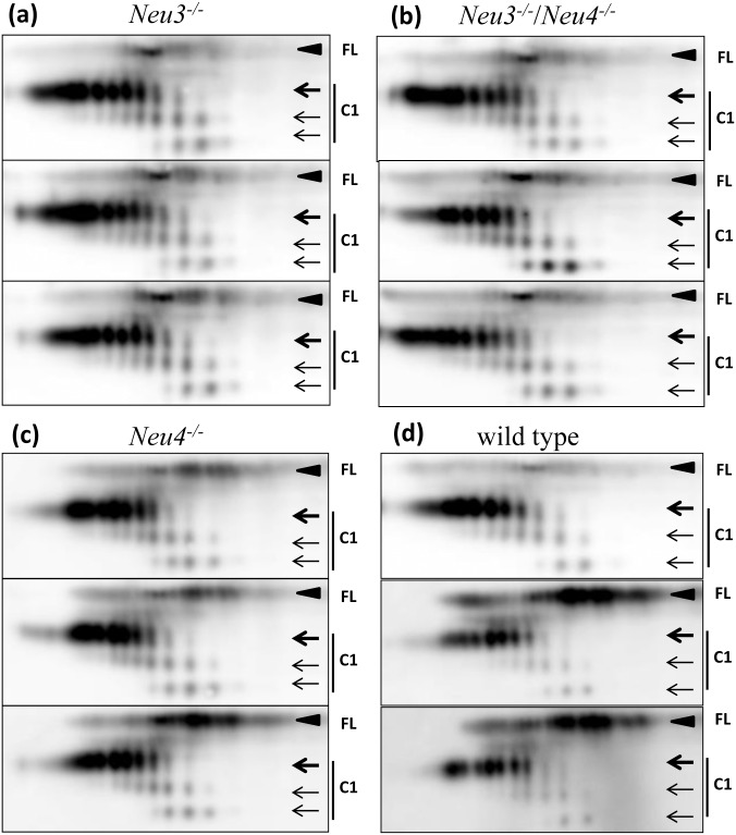
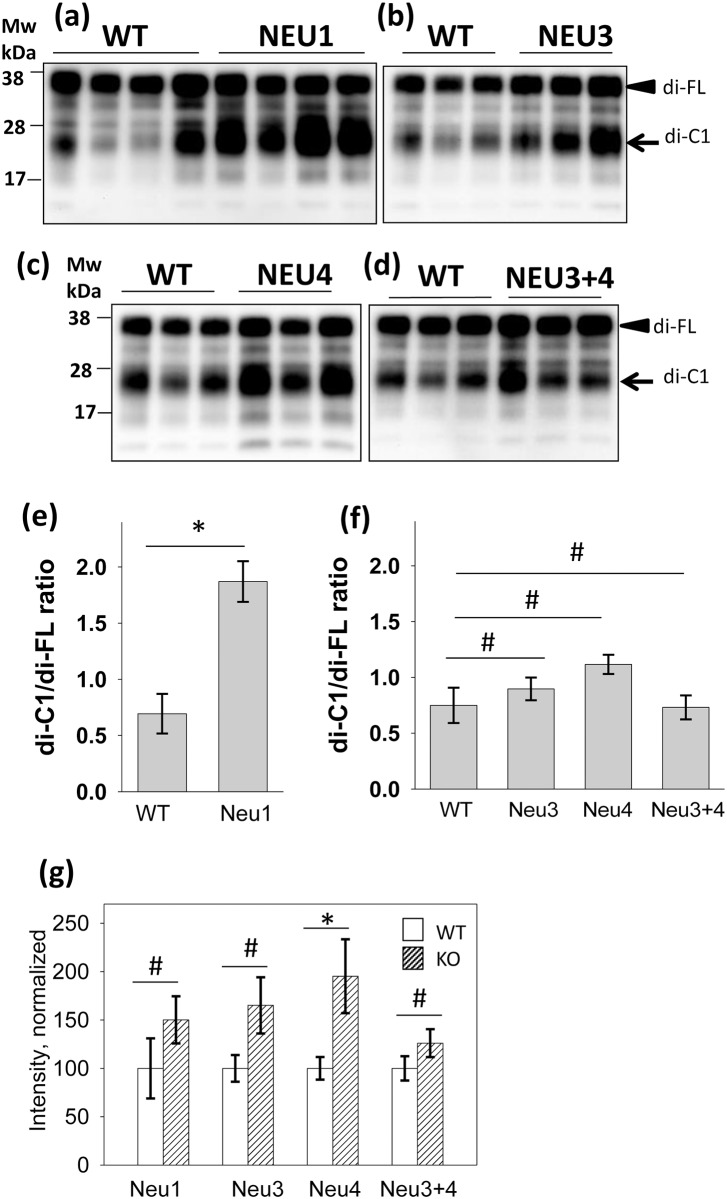
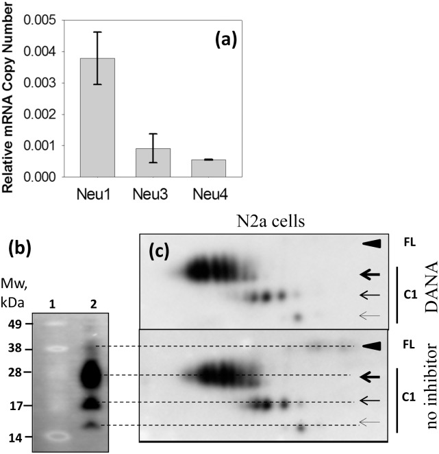
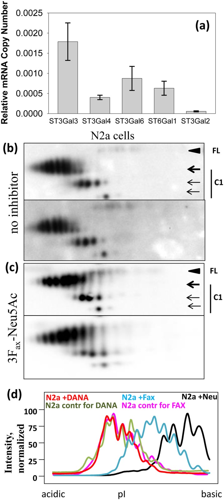
The central molecular event underlying prion diseases involves conformational change of the cellular form of the prion protein (PrPC), which is a sialoglycoprotein, into the disease-associated, transmissible form denoted PrPSc. Recent studies revealed a correlation between the sialylation status of PrPSc and incubation time to disease and introduced a new hypothesis that progression of prion diseases could be controlled or reversed by altering the sialylation level of PrPC. Of the four known mammalian sialidases, the enzymes that cleave off sialic acid residues, only NEU1, NEU3 and NEU4 are expressed in the brain. To test whether cellular sialidases control the steady-state sialylation level of PrPC and to identify the putative sialidase responsible for desialylating PrPC, we analyzed brain-derived PrPC from knockout mice deficient in Neu1, Neu3, Neu4, or from Neu3/Neu4 double knockouts. Surprisingly, no differences in the sialylation of PrPC or its proteolytic product C1 were noticed in any of the knockout mice tested as compared to the age-matched controls. However, significantly higher amounts of the C1 fragment relative to full-length PrPC were detected in the brains of Neu1 knockout mice as compared to WT mice or to the other knockout mice. Additional experiments revealed that in neuroblastoma cell line the sialylation pattern of C1 could be changed by an inhibitor of sialylatransferases. In summary, this study suggests that targeting cellular sialidases is apparently not the correct strategy for altering the sialylation levels of PrPC, whereas modulating the activity of sialylatransferases might offer a more promising approach. Our findings also suggest that catabolism of PrPC involves its α-cleavage followed by desialylation of the resulting C1 fragments by NEU1 and consequent fast degradation of the desialylated products.
朊病毒疾病的核心分子事件涉及朊病毒蛋白(PrPC)的细胞形式(一种唾液酸糖蛋白)构象转变为与疾病相关的、可传播的形式PrPSc。最近的研究揭示了PrPSc的唾液酸化状态与疾病潜伏期之间的相关性,并提出了一个新的假说,即通过改变PrPC的唾液酸化水平可以控制或逆转朊病毒疾病的进展。在四种已知的哺乳动物唾液酸酶(即切割去除唾液酸残基的酶)中,只有NEU1、NEU3和NEU4在大脑中表达。为了测试细胞唾液酸酶是否控制PrPC的稳态唾液酸化水平,并确定负责使PrPC去唾液酸化的假定唾液酸酶,我们分析了来自Neu1、Neu3、Neu4基因敲除小鼠或Neu3/Neu4双基因敲除小鼠脑源性PrPC。令人惊讶的是,与年龄匹配的对照相比,在所测试的任何基因敲除小鼠中,均未发现PrPC及其蛋白水解产物C1的唾液酸化有差异。然而,与野生型小鼠或其他基因敲除小鼠相比,在Neu1基因敲除小鼠的大脑中检测到相对于全长PrPC而言显著更多的C1片段。额外的实验表明,在神经母细胞瘤细胞系中,C1的唾液酸化模式可被唾液酸转移酶抑制剂改变。总之,本研究表明,靶向细胞唾液酸酶显然不是改变PrPC唾液酸化水平的正确策略,而调节唾液酸转移酶的活性可能提供一种更有前景的方法。我们的研究结果还表明,PrPC的分解代谢涉及其α-切割,随后由NEU1对产生的C1片段进行去唾液酸化,并导致去唾液酸化产物的快速降解。