Stec Małgorzata, Szatanek Rafał, Baj-Krzyworzeka Monika, Baran Jarosław, Zembala Maria, Barbasz Jakub, Waligórska Agnieszka, Dobrucki Jurek W, Mytar Bożenna, Szczepanik Antoni, Siedlar Maciej, Drabik Grażyna, Urbanowicz Barbara, Zembala Marek
Department of Clinical Immunology and Transplantology, Jagiellonian University Medical College, Wielicka 265 Str., 30-663, Kraków, Poland.
Institute of Catalysis and Surface Chemistry, Polish Academy of Sciences, Kraków, Poland.
J Transl Med. 2015 Dec 1;13:376. doi: 10.1186/s12967-015-0737-0.
Tumour cells release membrane micro(nano)fragments called tumour-derived microvesicles (TMV) that are believed to play an important role in cancer progression. TMV suppress/modify antitumour response of the host, but there is also some evidence for their direct interaction with cancer cells. In cancer patients TMV are present in body fluid and tumour microenvironment. The present study aimed at characterization of whole types/subpopulations, but not only exosomes, of TMV from newly established gastric cancer cell line (called GC1415) and to define their interactions with autologous cells.
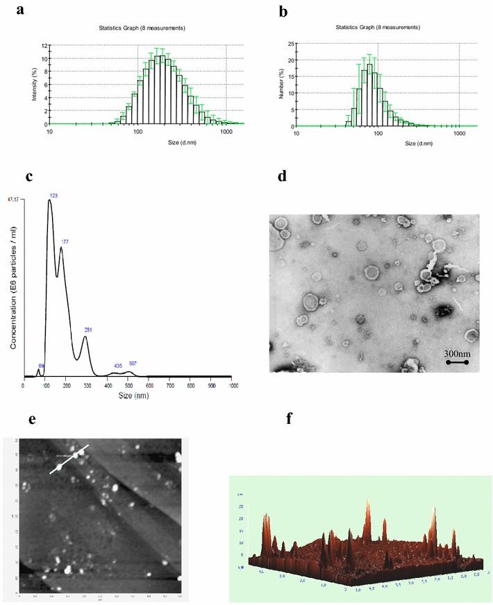
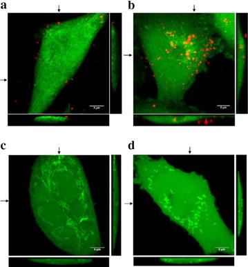
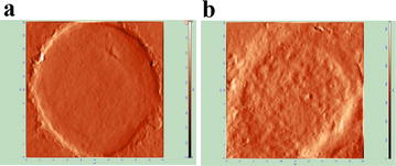
TMV were isolated from cell cultures supernatants by centrifugation at 50,000×g and their phenotype was determined by flow cytometry. The size of TMV was analysed by dynamic light scattering and nanoparticle tracking analysis, while morphology by transmission electron microscopy and atomic force microscopy. Interactions of TMV with cancer cells were visualized using fluorescence-activated cell sorter, confocal and atomic force microscopy, biological effects by xenografts in NOD SCID mice.
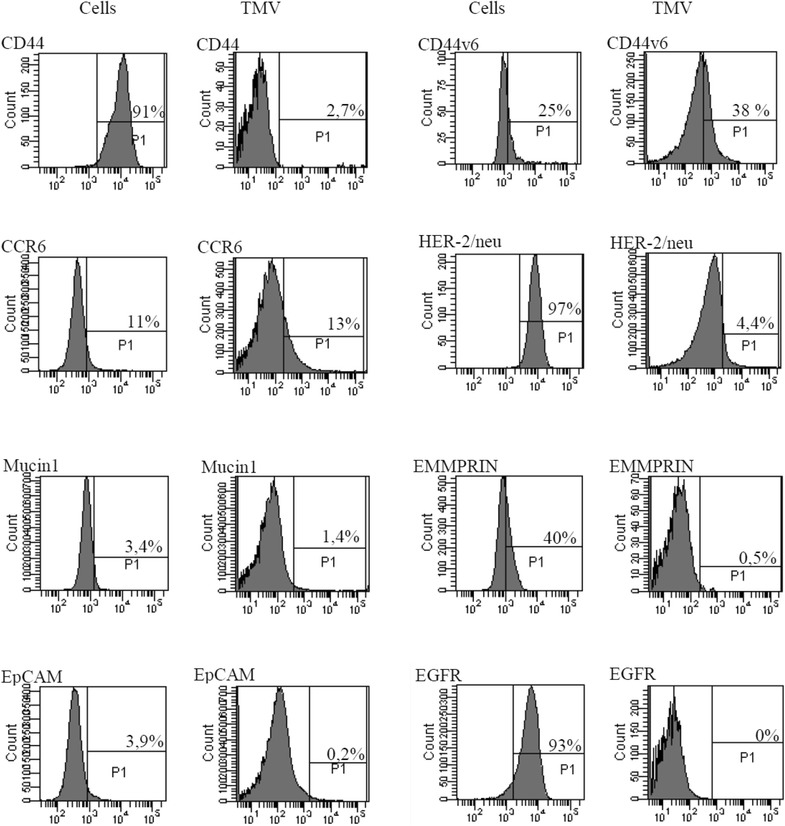
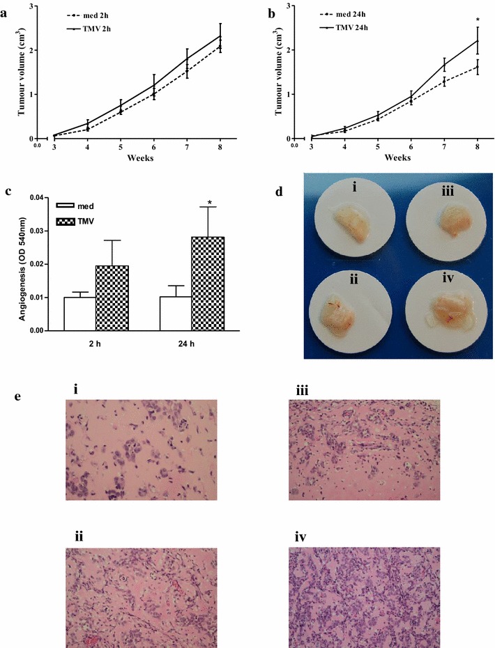
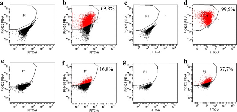
Isolated TMV showed expression of CD44H, CD44v6 (hyaluronian receptors), CCR6 (chemokine receptor) and HER-2/neu molecules, exhibited different shapes and sizes (range 60-900 nm, highest frequency of particles with size range of 80-120 nm). TMV attached to autologous cancer cells within 2 h and then were internalized by them at 24 h. CD44H, CD44v6 and CCR6 molecules may play a role in attachment of TMV to cancer cells, while HER-2 associated with CD24 be involved in promoting cancer cells growth. Pre-exposure of cancer cells to TMV resulted in enhancement of tumour growth and cancer cell-induced angiogenesis in NOD SCID mice model.
TMV interact directly with cancer cells serving as macro-messengers and molecular cargo transfer between gastric cancer cells resulting in enhancement of tumour growth. TMV should be considered in future as target of anticancer therapy.
肿瘤细胞释放被称为肿瘤衍生微泡(TMV)的膜微(纳)片段,据信其在癌症进展中起重要作用。TMV抑制/改变宿主的抗肿瘤反应,但也有一些证据表明它们与癌细胞直接相互作用。在癌症患者中,TMV存在于体液和肿瘤微环境中。本研究旨在对新建立的胃癌细胞系(称为GC1415)的TMV的所有类型/亚群进行表征,而不仅仅是外泌体,并确定它们与自体细胞的相互作用。
通过50,000×g离心从细胞培养上清液中分离TMV,并通过流式细胞术确定其表型。通过动态光散射和纳米颗粒跟踪分析来分析TMV的大小,而通过透射电子显微镜和原子力显微镜来观察形态。使用荧光激活细胞分选仪、共聚焦显微镜和原子力显微镜观察TMV与癌细胞的相互作用,通过在NOD SCID小鼠中进行异种移植来观察生物学效应。
分离出的TMV显示出CD44H、CD44v6(透明质酸受体)、CCR6(趋化因子受体)和HER-2/neu分子的表达,呈现出不同的形状和大小(范围为60-900nm,大小范围为80-120nm的颗粒频率最高)。TMV在两小时内附着于自体癌细胞,然后在24小时时被它们内化。CD44H、CD44v6和CCR6分子可能在TMV与癌细胞的附着中起作用,而与CD24相关的HER-2参与促进癌细胞生长。在NOD SCID小鼠模型中,使癌细胞预先暴露于TMV导致肿瘤生长和癌细胞诱导的血管生成增强。
TMV作为大分子信使直接与癌细胞相互作用,并在胃癌细胞之间进行分子货物转移,从而导致肿瘤生长增强。未来应将TMV视为抗癌治疗的靶点。