Yonezawa Satoshi, Shigematsu Momoko, Hirata Kazuto, Hayashi Kensuke
Department of Materials and Life Sciences, Faculty of Science and Technology, Sophia University , 7-1 Kioicho, Chiyoda-ku, Tokyo, Japan.
Acta Histochem Cytochem. 2015 Oct 29;48(5):145-52. doi: 10.1267/ahc.15023. Epub 2015 Oct 21.
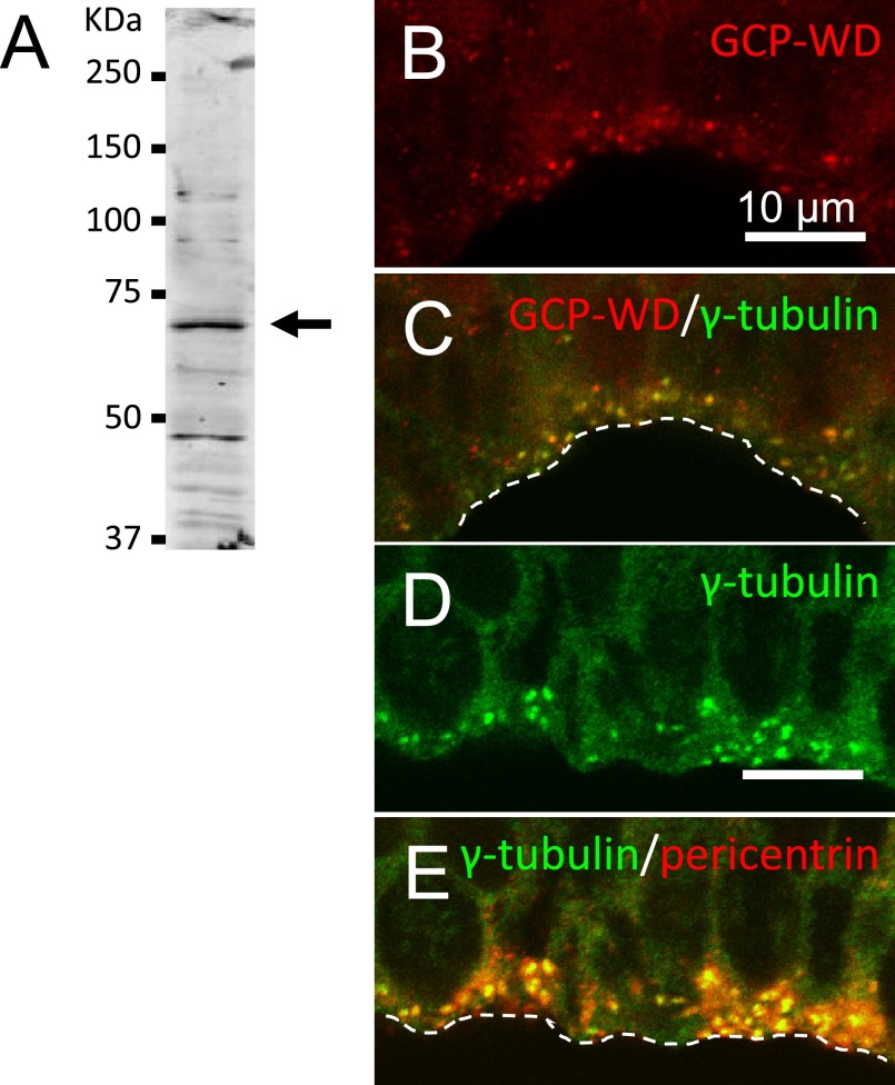
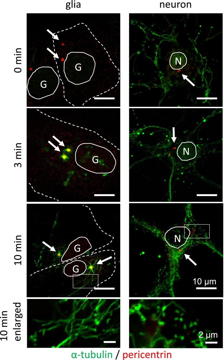
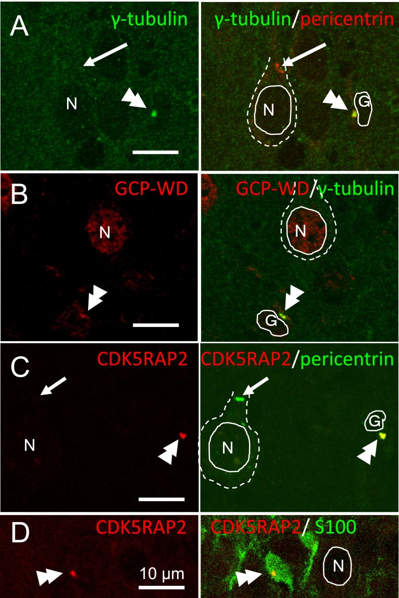
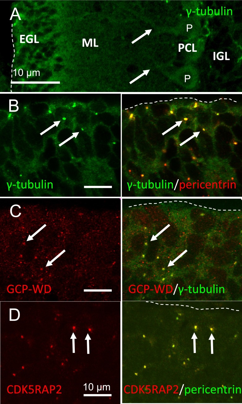
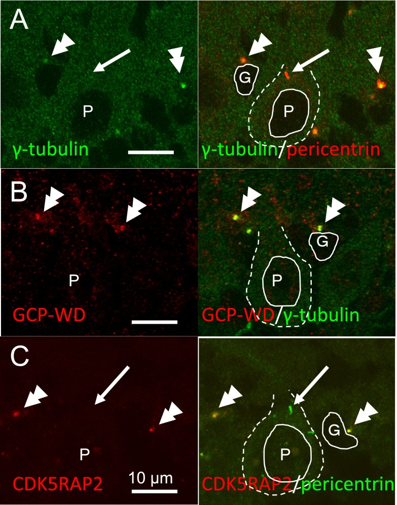
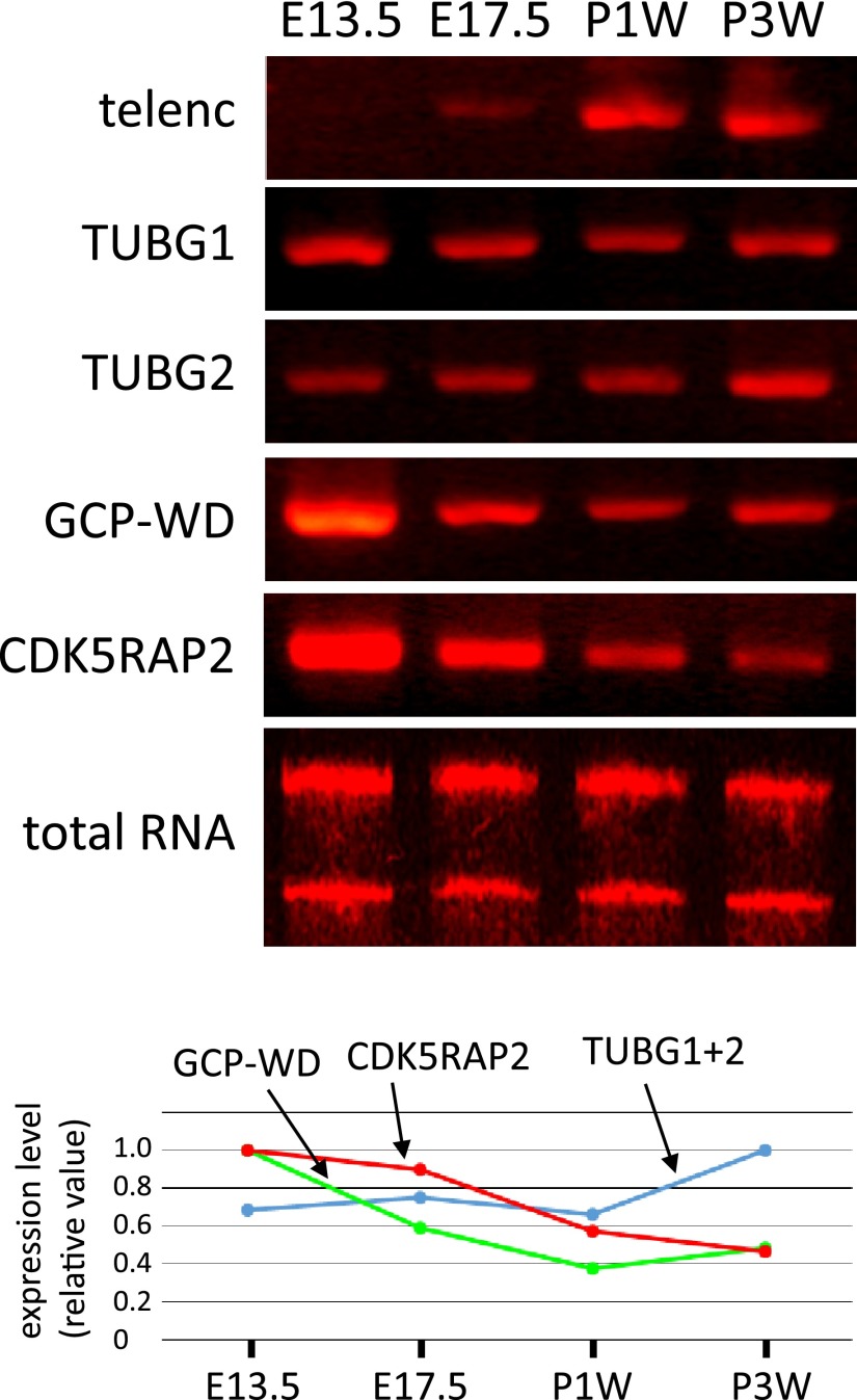
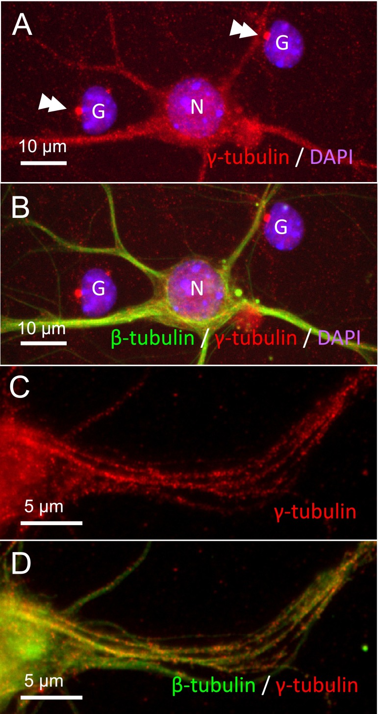
It has been recently reported that the centrosome of neurons does not have microtubule nucleating activity. Microtubule nucleation requires γ-tubulin as well as its recruiting proteins, GCP-WD/NEDD1 and CDK5RAP2 that anchor γ-tubulin to the centrosome. Change in the localization of these proteins during in vivo development of brain, however, has not been well examined. In this study we investigate the localization of γ-tubulin, GCP-WD and CDK5RAP2 in developing cerebral and cerebellar cortex with immunofluorescence. We found that γ-tubulin and its recruiting proteins were localized at centrosomes of immature neurons, while they were lost at centrosomes in mature neurons. This indicated that the loss of microtubule nucleating activity at the centrosome of neurons is due to the loss of γ-tubulin-recruiting proteins from the centrosome. RT-PCR analysis revealed that these proteins are still expressed after birth, suggesting that they have a role in microtubule generation in cell body and dendrites of mature neurons. Microtubule regrowth experiments on cultured mature neurons showed that microtubules are nucleated not at the centrosome but within dendrites. These data indicated the translocation of microtubule-organizing activity from the centrosome to dendrites during maturation of neurons, which would explain the mixed polarity of microtubules in dendrites.
最近有报道称,神经元的中心体不具有微管成核活性。微管成核需要γ-微管蛋白及其招募蛋白GCP-WD/NEDD1和CDK5RAP2,这些蛋白将γ-微管蛋白锚定在中心体上。然而,在大脑的体内发育过程中,这些蛋白的定位变化尚未得到充分研究。在本研究中,我们用免疫荧光法研究了γ-微管蛋白、GCP-WD和CDK5RAP2在发育中的大脑皮层和小脑皮层中的定位。我们发现,γ-微管蛋白及其招募蛋白定位于未成熟神经元的中心体,而在成熟神经元的中心体中则消失。这表明神经元中心体微管成核活性的丧失是由于中心体中γ-微管蛋白招募蛋白的丧失。逆转录聚合酶链反应(RT-PCR)分析显示,这些蛋白在出生后仍有表达,表明它们在成熟神经元的细胞体和树突中的微管生成中起作用。对培养的成熟神经元进行的微管再生实验表明,微管不是在中心体而是在树突内成核。这些数据表明,在神经元成熟过程中,微管组织活性从中心体转移到树突,这可以解释树突中微管的混合极性。