Koch Karl-Wilhelm, Dell'Orco Daniele
Department of Neurosciences, Biochemistry Group, University of Oldenburg Oldenburg, Germany.
Department of Neurological, Biomedical and Movement Sciences, Section of Biological Chemistry and Center for BioMedical Computing (CBMC), University of Verona Verona, Italy.
Front Mol Neurosci. 2015 Nov 17;8:67. doi: 10.3389/fnmol.2015.00067. eCollection 2015.
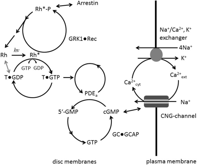
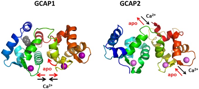
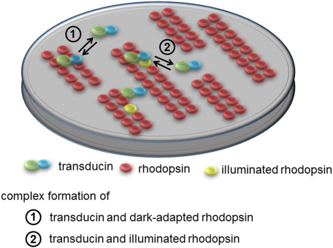
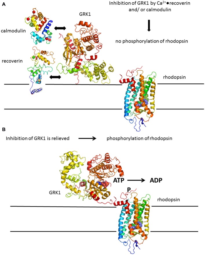
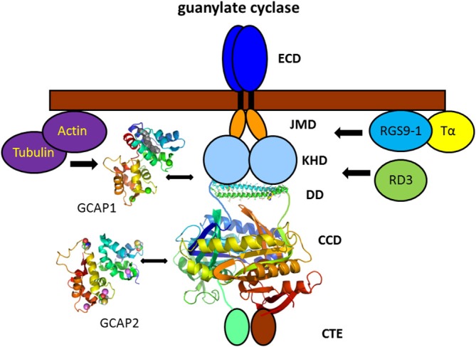
Vertebrate photoreceptor cells are exquisite light detectors operating under very dim and bright illumination. The photoexcitation and adaptation machinery in photoreceptor cells consists of protein complexes that can form highly ordered supramolecular structures and control the homeostasis and mutual dependence of the secondary messengers cyclic guanosine monophosphate (cGMP) and Ca(2+). The visual pigment in rod photoreceptors, the G protein-coupled receptor rhodopsin is organized in tracks of dimers thereby providing a signaling platform for the dynamic scaffolding of the G protein transducin. Illuminated rhodopsin is turned off by phosphorylation catalyzed by rhodopsin kinase (GRK1) under control of Ca(2+)-recoverin. The GRK1 protein complex partly assembles in lipid raft structures, where shutting off rhodopsin seems to be more effective. Re-synthesis of cGMP is another crucial step in the recovery of the photoresponse after illumination. It is catalyzed by membrane bound sensory guanylate cyclases (GCs) and is regulated by specific neuronal Ca(2+)-sensor proteins called guanylate cyclase-activating proteins (GCAPs). At least one GC (ROS-GC1) was shown to be part of a multiprotein complex having strong interactions with the cytoskeleton and being controlled in a multimodal Ca(2+)-dependent fashion. The final target of the cGMP signaling cascade is a cyclic nucleotide-gated (CNG) channel that is a hetero-oligomeric protein located in the plasma membrane and interacting with accessory proteins in highly organized microdomains. We summarize results and interpretations of findings related to the inhomogeneous organization of signaling units in photoreceptor outer segments.
脊椎动物的光感受器细胞是在非常昏暗和明亮的光照条件下工作的精密光探测器。光感受器细胞中的光激发和适应机制由蛋白质复合物组成,这些复合物可以形成高度有序的超分子结构,并控制二级信使环鸟苷单磷酸(cGMP)和Ca(2+)的稳态及相互依赖性。视杆光感受器中的视觉色素,即G蛋白偶联受体视紫红质,以二聚体形式排列成轨道,从而为G蛋白转导素的动态支架提供了一个信号平台。在Ca(2+) - 恢复蛋白的控制下,视紫红质激酶(GRK1)催化的磷酸化作用会使被光照激活的视紫红质失活。GRK1蛋白复合物部分组装在脂筏结构中,在那里使视紫红质失活似乎更有效。cGMP的重新合成是光照后光反应恢复的另一个关键步骤。它由膜结合的感觉鸟苷酸环化酶(GCs)催化,并由称为鸟苷酸环化酶激活蛋白(GCAPs)的特定神经元Ca(2+) - 传感器蛋白调节。至少有一种GC(ROS - GC1)被证明是多蛋白复合物的一部分,该复合物与细胞骨架有强烈相互作用,并以多模式Ca(2+)依赖性方式受到控制。cGMP信号级联的最终靶点是一个环核苷酸门控(CNG)通道,它是一种位于质膜中的异源寡聚蛋白,在高度组织化的微结构域中与辅助蛋白相互作用。我们总结了与光感受器外段信号单元不均匀组织相关的研究结果和解释。