Wang Xiaojie, Pettersson David R, Studholme Colin, Kroenke Christopher D
Division of Neuroscience, Oregon National Primate Research Center, Oregon Health & Science University Beaverton, OR, USA.
Department of Radiology, Oregon Health & Science University Portland, OR, USA.
Front Neuroanat. 2015 Nov 24;9:147. doi: 10.3389/fnana.2015.00147. eCollection 2015.
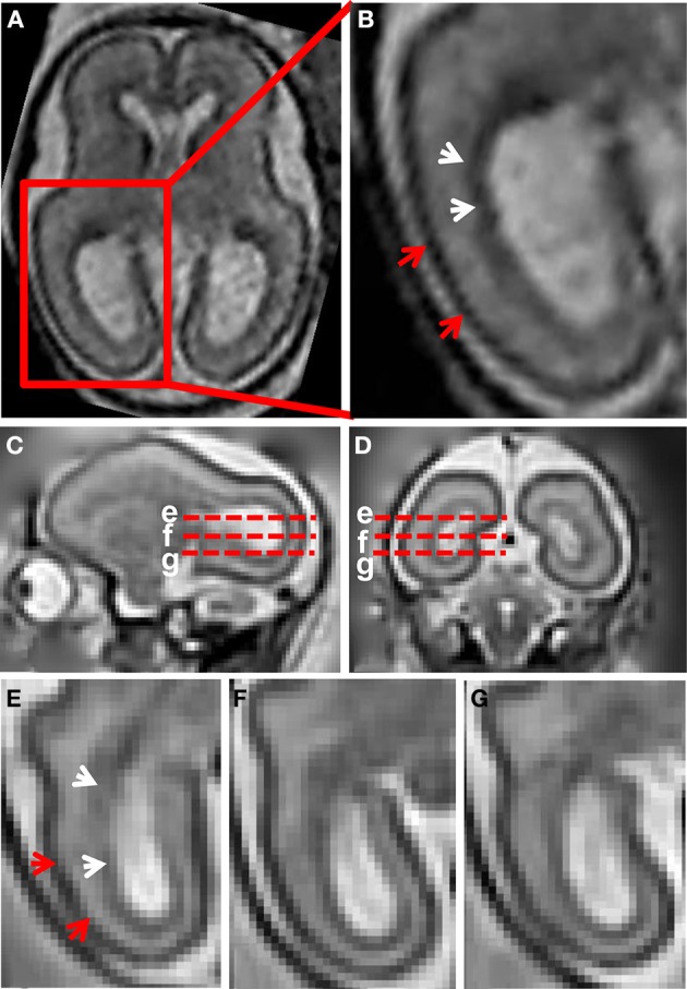
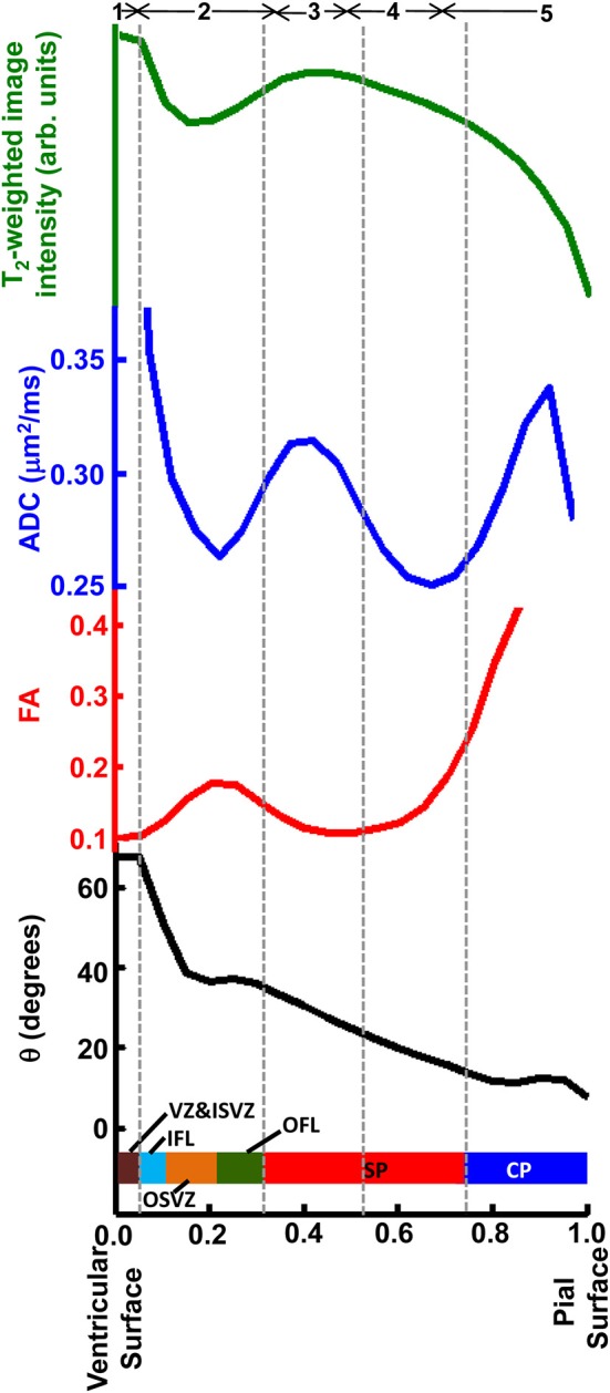
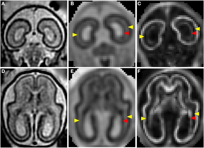
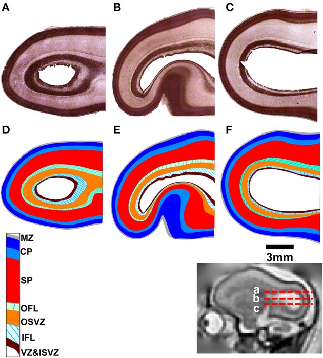
Distinct populations of progenitor and postmitotic neural and glial cells are stratified in the fetal primate brain across developmentally transient tissue zones between the ventricular and pial surfaces. These zones were originally identified by light microscopy. However, it has subsequently been shown that various forms of magnetic resonance image (MRI) contrast can be used to distinguish layers of developing neural tissue in ex vivo, as well as in vivo (including in utero) conditions. Here we compare mid-gestation rhesus macaque tissue zones identified using histological techniques to ex vivo as well as in utero MRI performed on the same brains. These data are compared to mid-gestation fetal human brain MRI results, obtained in utero. We observe strong similarity between MRI contrast in vivo and post mortem, which facilitates interpretation of in utero images based on the histological characterization performed here. Additionally, we observe differential correspondence between the various forms of ex vivo MRI contrast and microscopy data, with maps of the water apparent diffusion coefficient providing the closest match to histologically-identified lamina of the nonhuman primate brain. Examination of histology and post mortem MRI helps to provide a better understanding of cytoarchitectrual characteristics that give rise to in utero MRI contrast.
在灵长类胎儿大脑中,祖细胞以及有丝分裂后的神经细胞和神经胶质细胞的不同群体,在脑室表面和软脑膜表面之间发育短暂的组织区域内呈分层分布。这些区域最初是通过光学显微镜识别的。然而,随后的研究表明,各种形式的磁共振成像(MRI)造影可用于区分离体以及活体(包括子宫内)条件下发育中的神经组织层。在此,我们将使用组织学技术识别的妊娠中期恒河猴组织区域,与在同一大脑上进行的离体及子宫内MRI结果进行比较。这些数据与在子宫内获得的妊娠中期胎儿人脑MRI结果进行比较。我们观察到活体和死后MRI造影之间有很强的相似性,这有助于根据此处进行的组织学特征解释子宫内图像。此外,我们观察到各种离体MRI造影形式与显微镜数据之间存在差异对应关系,其中水表观扩散系数图与非人类灵长类动物大脑组织学鉴定的层最为匹配。对组织学和死后MRI的检查有助于更好地理解产生子宫内MRI造影的细胞结构特征。