Suckling John, Simas Tiago, Chattopadhyay Shayanti, Tait Roger, Su Li, Williams Guy, Rowe James B, O'Brien John T
Department of Psychiatry, University of Cambridge Cambridge, UK ; Cambridge and Peterborough Foundation NHS Trust Cambridge, UK ; MRC/Wellcome Trust Behavioural and Clinical Neuroscience Institute, University of Cambridge Cambridge, UK.
Department of Psychiatry, University of Cambridge Cambridge, UK.
Front Comput Neurosci. 2015 Nov 18;9:140. doi: 10.3389/fncom.2015.00140. eCollection 2015.
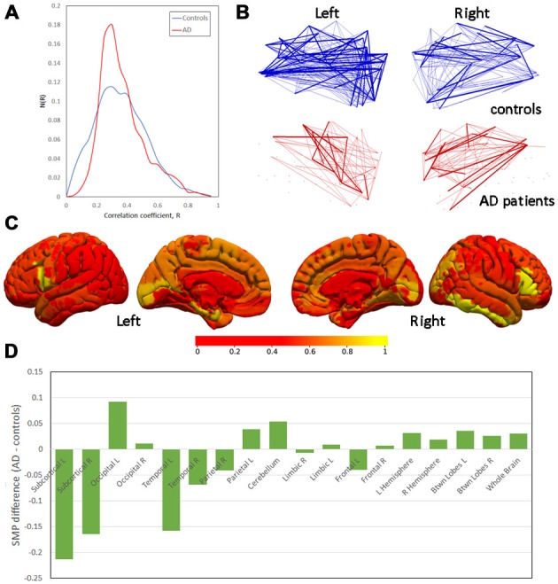
Neuroimaging has been successful in characterizing the pattern of cerebral atrophy that accompanies the progression of Alzheimer's disease (AD). Examination of functional connectivity, the strength of signal synchronicity between brain regions, has gathered pace as another way of understanding changes to the brain that are associated with AD. It appears to have good sensitivity and detect effects that precede cognitive decline, and thus offers the possibility to understand the neurobiology of the disease in its earliest phases. However, functional connectivity analyzes to date generally consider only the strongest connections, with weaker links ignored. This proof-of-concept study compared patients with mild-to-moderate AD (N = 11) and matched control individuals (N = 12) based on functional connectivities derived from blood-oxygenation level dependent (BOLD) sensitive functional MRI acquired during resting wakefulness. All positive connectivities irrespective of their strength were included. Transitive closures of the resulting connectome were calculated that classified connections as either direct or indirect. Between-group differences in the proportion of indirect paths were observed. In AD, there was broadly increased indirect connectivity across greater spatial distances. Furthermore, the indirect pathways in AD had greater between-subject topological variance than controls. The prevailing characterization of AD as being a disconnection syndrome is refined by the observation that direct links between regions that are impaired are perhaps replaced by an increase in indirect functional pathways that is only detectable through inclusion of connections across the entire range of connection strengths.
神经影像学已成功地描绘出伴随阿尔茨海默病(AD)进展的脑萎缩模式。作为理解与AD相关的大脑变化的另一种方式,对功能连接性(脑区之间信号同步性的强度)的研究也在加快步伐。它似乎具有良好的敏感性,能检测到认知衰退之前的影响,因此提供了在疾病最早阶段了解其神经生物学的可能性。然而,迄今为止的功能连接性分析通常只考虑最强的连接,而忽略了较弱的连接。这项概念验证研究基于静息清醒状态下采集的血氧水平依赖(BOLD)敏感功能磁共振成像得出的功能连接性,比较了轻度至中度AD患者(N = 11)和匹配的对照个体(N = 12)。所有正连接性,无论其强度如何,均被纳入。计算了所得脑连接组的传递闭包,将连接分类为直接或间接。观察到组间间接路径比例的差异。在AD中,跨更大空间距离的间接连接普遍增加。此外,AD中的间接通路在个体间的拓扑差异比对照组更大。AD作为一种失连接综合征的普遍特征通过以下观察得到完善:受损区域之间的直接连接可能被间接功能通路的增加所取代,而这种增加只有通过纳入整个连接强度范围内的连接才能检测到。