Hou Shaoping, Carson David M, Wu Di, Klaw Michelle C, Houlé John D, Tom Veronica J
Spinal Cord Research Center, Department of Neurobiology & Anatomy, Drexel University College of Medicine, Philadelphia, PA 19129, United States.
Spinal Cord Research Center, Department of Neurobiology & Anatomy, Drexel University College of Medicine, Philadelphia, PA 19129, United States.
Exp Neurol. 2016 Nov;285(Pt B):136-146. doi: 10.1016/j.expneurol.2015.12.001. Epub 2015 Dec 2.
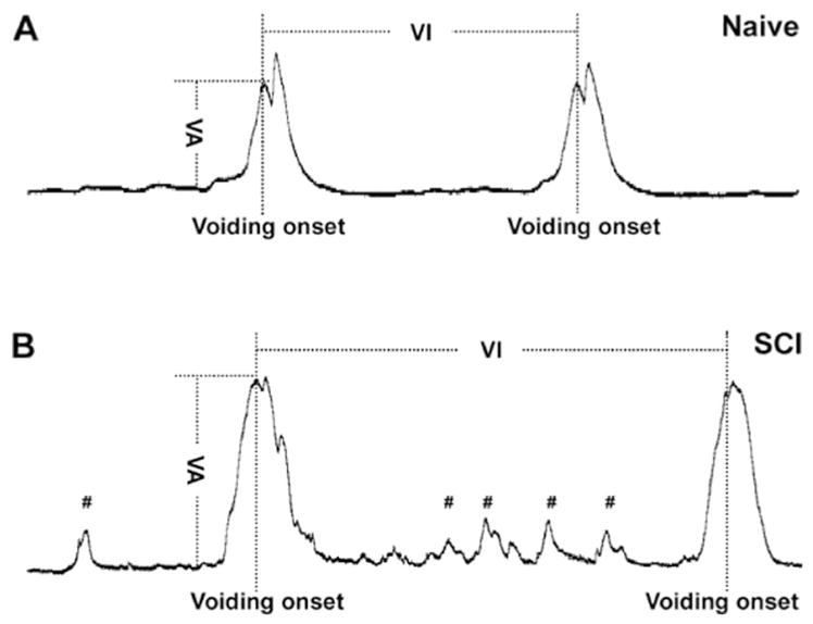
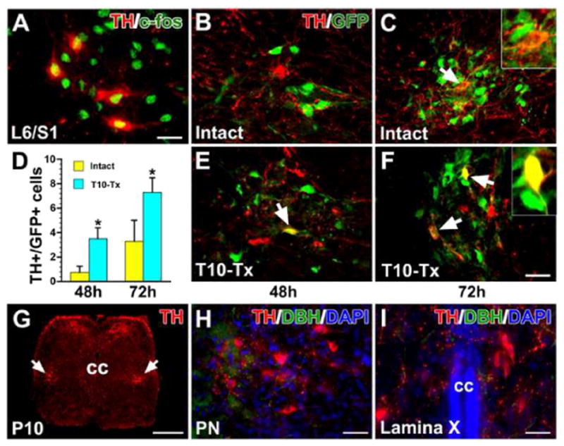
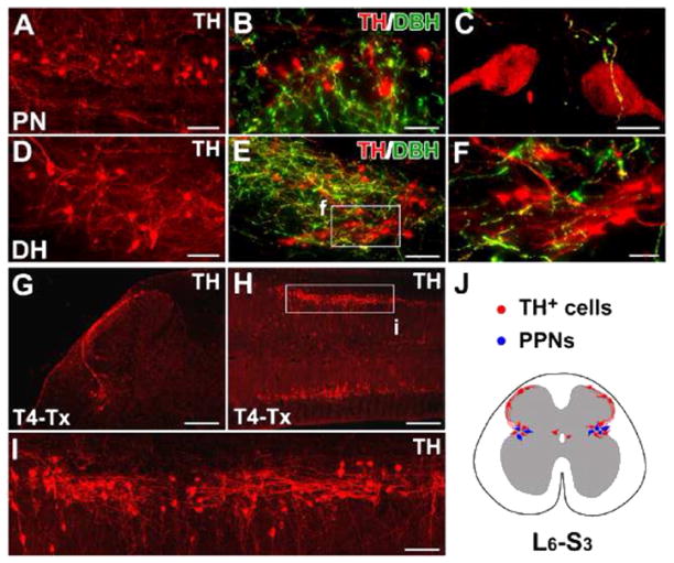
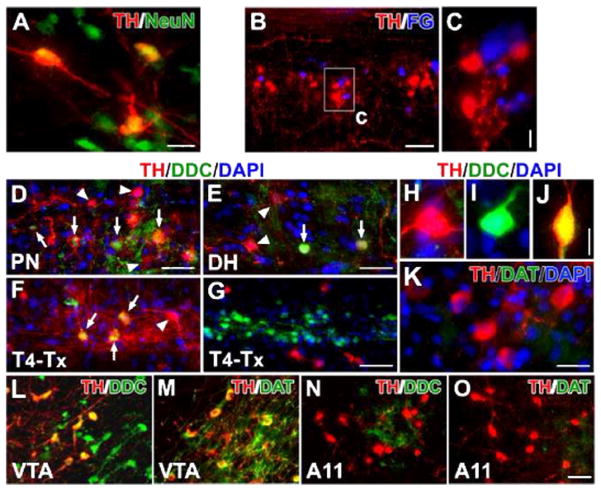
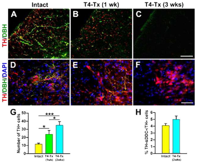
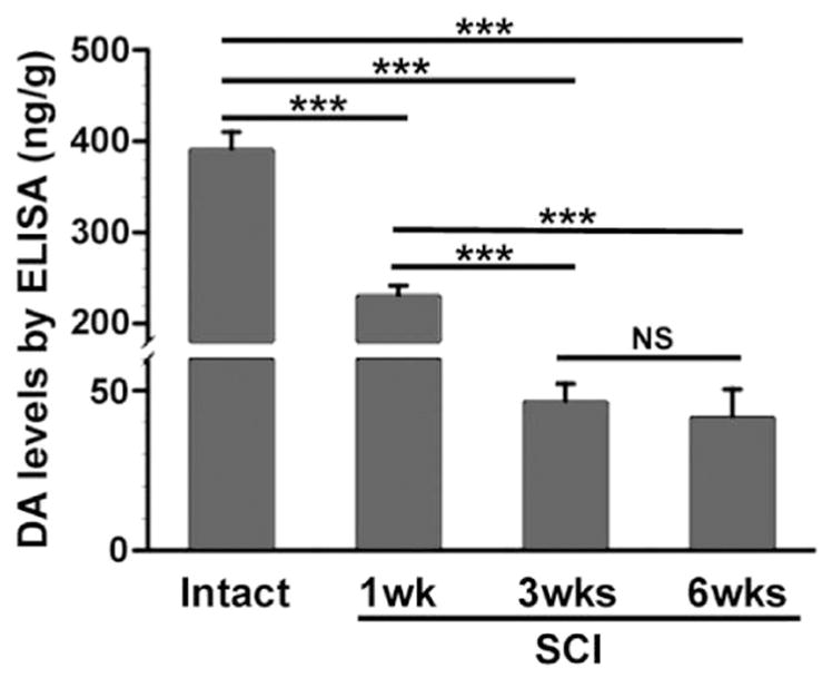
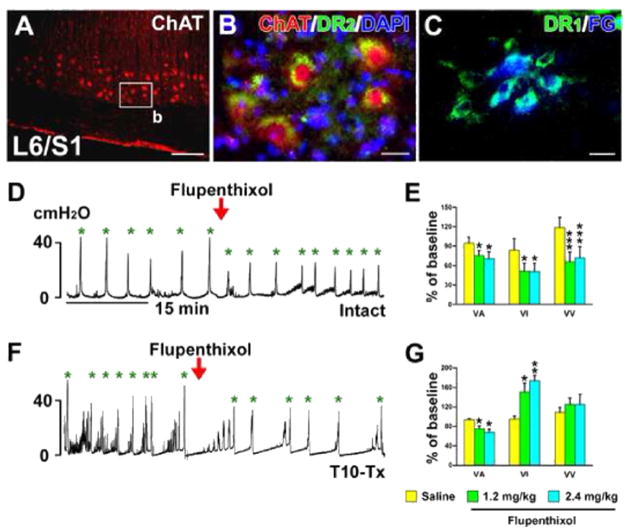
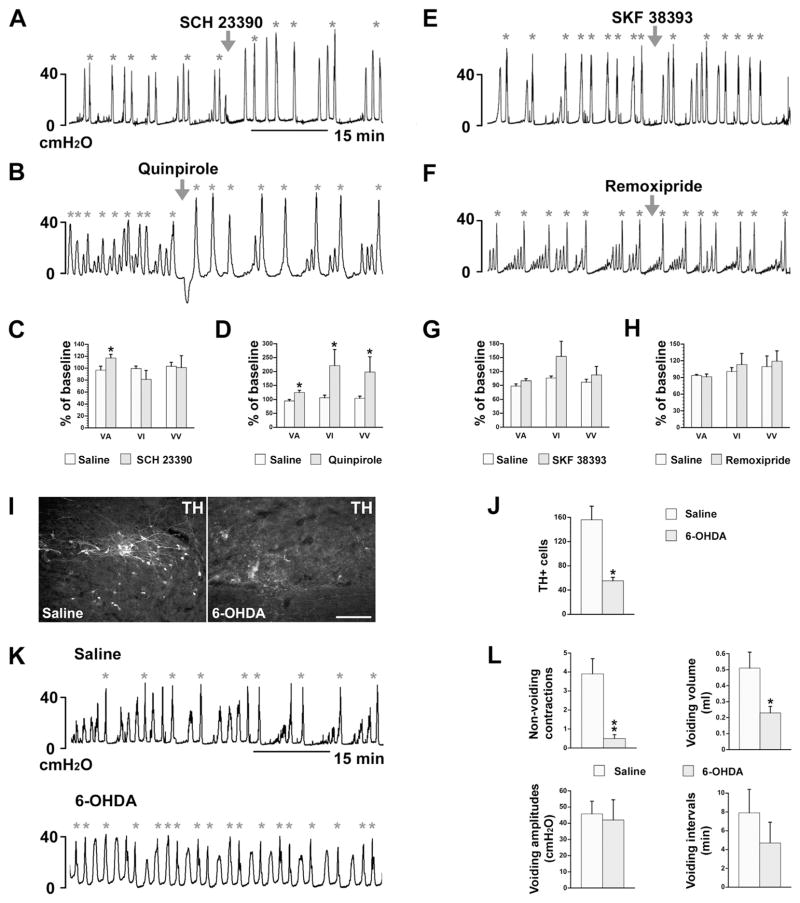
Dopamine (DA) neurons in the mammalian central nervous system are thought to be restricted to the brain. DA-mediated regulation of urinary activity is considered to occur through an interaction between midbrain DA neurons and the pontine micturition center. Here we show that DA is produced in the rat spinal cord and modulates the bladder reflex. We observed numerous tyrosine hydroxylase (TH) neurons in the autonomic nuclei and superficial dorsal horn in L6-S3 spinal segments. These neurons are dopamine-β-hydroxylase (DBH) and some contain detectable dopamine decarboxylase (DDC), suggesting their capacity to produce DA. Interestingly, following a complete thoracic spinal cord injury (SCI) to interrupt supraspinal projections, more TH neurons emerged in the lumbosacral spinal cord, coincident with a sustained, low level of DA expression there and a partially recovered micturition reflex. Non-selective blockade of spinal DA receptors reduced bladder activity whereas activation of spinal D-like receptors increased bladder activity and facilitated voiding. Additionally, depletion of lumbosacral TH neurons with 6-hydroxydopamine (6-OHDA) decreased bladder non-voiding contractions and voiding efficiency. Furthermore, injecting the transsynaptic neuronal tracer pseudorabies virus (PRV) into the bladder detrusor labeled TH cells in the lumbosacral cord, confirming their involvement in spinal micturition reflex circuits. These results illustrate that DA is synthesized in the rat spinal cord; plasticity of lumbosacral TH neurons following SCI may contribute to DA expression and modulate the spinal bladder reflex. Thus, spinally-derived DA and receptors could be a novel therapeutic target to improve micturition recovery after SCI.
哺乳动物中枢神经系统中的多巴胺(DA)神经元被认为局限于大脑。DA介导的泌尿活动调节被认为是通过中脑DA神经元与脑桥排尿中枢之间的相互作用来实现的。在此,我们表明DA在大鼠脊髓中产生并调节膀胱反射。我们在L6 - S3脊髓节段的自主神经核和浅表背角中观察到大量酪氨酸羟化酶(TH)神经元。这些神经元是多巴胺-β-羟化酶(DBH),并且一些含有可检测到的多巴胺脱羧酶(DDC),表明它们具有产生DA的能力。有趣的是,在完全性胸段脊髓损伤(SCI)以中断脊髓上投射后,腰骶部脊髓中出现了更多的TH神经元,同时那里DA表达持续处于低水平,且排尿反射部分恢复。脊髓DA受体的非选择性阻断降低了膀胱活动,而脊髓D样受体的激活增加了膀胱活动并促进了排尿。此外,用6 - 羟基多巴胺(6 - OHDA)耗尽腰骶部TH神经元会降低膀胱非排尿收缩和排尿效率。此外,将跨突触神经元示踪剂伪狂犬病病毒(PRV)注入膀胱逼尿肌后,标记了腰骶部脊髓中的TH细胞,证实它们参与脊髓排尿反射回路。这些结果表明DA在大鼠脊髓中合成;SCI后腰骶部TH神经元的可塑性可能有助于DA表达并调节脊髓膀胱反射。因此,脊髓源性DA及其受体可能是改善SCI后排尿恢复的新治疗靶点。