• 文献检索
  • 文档翻译
  • 深度研究
  • 学术资讯
  • Suppr Zotero 插件Zotero 插件
  • 邀请有礼
  • 套餐&价格
  • 历史记录
应用&插件
Suppr Zotero 插件Zotero 插件浏览器插件Mac 客户端Windows 客户端微信小程序
定价
高级版会员购买积分包购买API积分包
服务
文献检索文档翻译深度研究API 文档MCP 服务
关于我们
关于 Suppr公司介绍联系我们用户协议隐私条款
关注我们

Suppr 超能文献

核心技术专利:CN118964589B侵权必究
粤ICP备2023148730 号-1Suppr @ 2026

文献检索

告别复杂PubMed语法,用中文像聊天一样搜索,搜遍4000万医学文献。AI智能推荐,让科研检索更轻松。

立即免费搜索

文件翻译

保留排版,准确专业,支持PDF/Word/PPT等文件格式,支持 12+语言互译。

免费翻译文档

深度研究

AI帮你快速写综述,25分钟生成高质量综述,智能提取关键信息,辅助科研写作。

立即免费体验

通过前囟对新生儿和婴儿中枢神经系统进行三维超声检查在评估颅内出血及其后果中的应用价值。

Usefulness of 3D sonography of the central nervous system in neonates and infants in the assessment of intracranial bleeding and its consequences when examined through the anterior fontanelle.

作者信息

Zielonka-Lamparska Edyta, Wieczorek Andrzej Paweł

机构信息

Zakład Radiologii Dziecięcej, Uniwersytet Medyczny w Lublinie, Lublin, Polska.

出版信息

J Ultrason. 2013 Dec;13(55):408-17. doi: 10.15557/JoU.2013.0043. Epub 2013 Dec 30.

DOI:10.15557/JoU.2013.0043
PMID:26675013
原文链接:https://pmc.ncbi.nlm.nih.gov/articles/PMC4579667/
Abstract

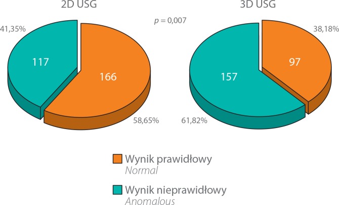
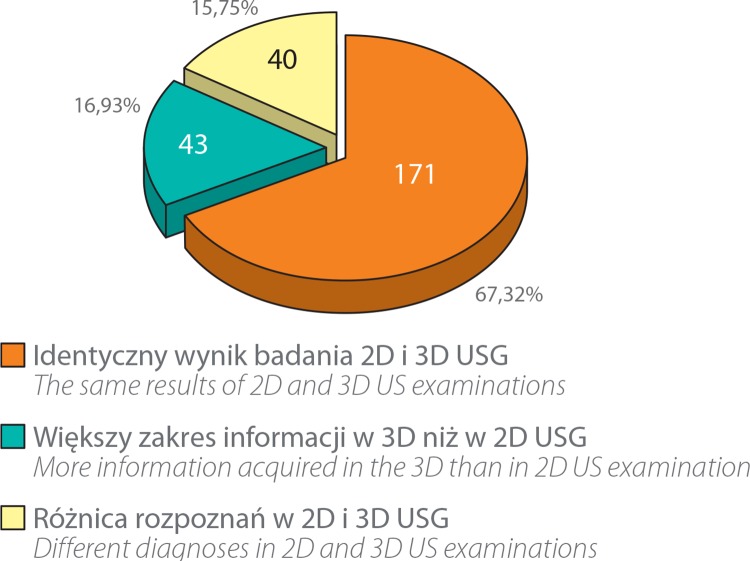
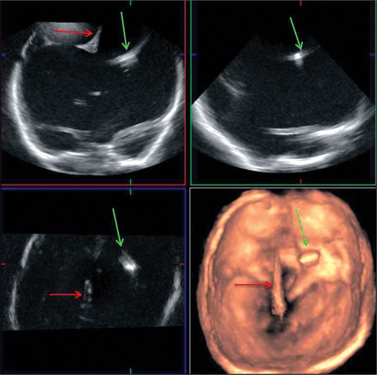
Due to the possibility to examine at the patient bedside or incubator, ultrasound imaging of the central nervous system, particularly through the anterior fontanelle, is the most common diagnostic examination performed in neonates and infants with neurological problems. Despite its common application, this method has certain limitations. These limitations are associated with cross-sections of the cerebral structures that can be obtained when examining through the anterior fontanelle. The aim of the paper was to assess the usefulness of three dimensional sonography of the central nervous system in neonates and infants in the assessment of intracranial bleeding and its consequences when examined through the anterior fontanelle. The study enrolled 283 patients treated at the Pediatric Teaching Hospital at the Medical University of Lublin in whom a transfontanelle cerebral examination was indicated. The two- and three-dimensional ultrasound examinations of the central nervous system were conducted in 283 patients aged from 1 day to 18 months (mean age: 2 months). 254 three-dimensional examinations were of diagnostic value. The number of detected pathological lesions was higher in a statistically significant way (p = 0.007) in the three-dimensional ultrasound examination. In the authors' own studies, the highest sensitivity and statistically significant superiority of the three-dimensional method over the two-dimensional one referred to detecting intraventricular or intracerebral hemorrhages. Novel techniques of ultrasound imaging, including the three-dimensional one, have undoubtedly increased the diagnostic possibilities of sonography and, at the same time, retained all its advantages.

摘要

由于能够在患者床边或保育箱中进行检查,中枢神经系统的超声成像,尤其是通过前囟进行的检查,是对有神经问题的新生儿和婴儿进行的最常见诊断检查。尽管其应用广泛,但这种方法有一定局限性。这些局限性与通过前囟检查时所能获得的脑结构横截面有关。本文的目的是评估新生儿和婴儿中枢神经系统三维超声检查在评估通过前囟检查时颅内出血及其后果方面的实用性。该研究纳入了283名在卢布林医科大学儿科教学医院接受治疗且需要进行经囟门脑部检查的患者。对283名年龄从1天到18个月(平均年龄:2个月)的患者进行了中枢神经系统的二维和三维超声检查。254次三维检查具有诊断价值。三维超声检查中检测到的病理性病变数量在统计学上有显著增加(p = 0.007)。在作者自己的研究中,三维方法在检测脑室内或脑内出血方面具有最高的敏感性,且在统计学上明显优于二维方法。超声成像的新技术,包括三维技术,无疑增加了超声检查的诊断可能性,同时保留了其所有优点。

https://cdn.ncbi.nlm.nih.gov/pmc/blobs/11e9/4579667/cdccd03748de/JoU-2013-0043-g005.jpg
https://cdn.ncbi.nlm.nih.gov/pmc/blobs/11e9/4579667/3895a72130ee/JoU-2013-0043-g001.jpg
https://cdn.ncbi.nlm.nih.gov/pmc/blobs/11e9/4579667/3bee71cf869d/JoU-2013-0043-g002.jpg
https://cdn.ncbi.nlm.nih.gov/pmc/blobs/11e9/4579667/d86b6fd71958/JoU-2013-0043-g003.jpg
https://cdn.ncbi.nlm.nih.gov/pmc/blobs/11e9/4579667/3ce9b53bfe4e/JoU-2013-0043-g004.jpg
https://cdn.ncbi.nlm.nih.gov/pmc/blobs/11e9/4579667/cdccd03748de/JoU-2013-0043-g005.jpg

相似文献

1
Usefulness of 3D sonography of the central nervous system in neonates and infants in the assessment of intracranial bleeding and its consequences when examined through the anterior fontanelle.通过前囟对新生儿和婴儿中枢神经系统进行三维超声检查在评估颅内出血及其后果中的应用价值。
J Ultrason. 2013 Dec;13(55):408-17. doi: 10.15557/JoU.2013.0043. Epub 2013 Dec 30.
2
Diagnosis of intraventricular hemorrhage in the newborn: value of sonography via the posterior fontanelle.新生儿脑室内出血的诊断:经后囟门超声检查的价值
AJR Am J Roentgenol. 1994 Oct;163(4):893-6. doi: 10.2214/ajr.163.4.8092030.
3
[Infratentorial pathology a pitfall for neonatal cerebral sonography: Additional sonographic imaging through the posterolateral fontanelle, a prospective study].[幕下病变:新生儿颅脑超声检查的一个陷阱——通过后外侧囟门的附加超声成像,一项前瞻性研究]
Klin Padiatr. 2002 Mar-Apr;214(2):76-8. doi: 10.1055/s-2002-25274.
4
Use of posterior fontanelle in the ultrasound diagnosis of intraventricular/periventricular hemorrhage.后囟门在脑室内/脑室周围出血超声诊断中的应用。
J Pediatr (Rio J). 2008 Nov-Dec;84(6):503-8. doi: 10.2223/JPED.1855.
5
Normal and abnormal development of the fetal anterior fontanelle: a three-dimensional ultrasound study.胎儿前囟门的正常与异常发育:一项三维超声研究
Ultrasound Obstet Gynecol. 2008 Nov;32(6):755-61. doi: 10.1002/uog.5368.
6
Three-dimensional transvaginal neurosonography of the fetal brain: 'navigating' in the volume scan.胎儿脑部的三维经阴道神经超声检查:在容积扫描中“导航”
Ultrasound Obstet Gynecol. 2000 Sep;16(4):307-13. doi: 10.1046/j.1469-0705.2000.00264.x.
7
Sonography of ventricular size and germinal matrix hemorrhage in premature infants.早产儿脑室大小及生发基质出血的超声检查
AJR Am J Roentgenol. 1980 Sep;135(3):559-64. doi: 10.2214/ajr.135.3.559.
8
A new pattern of cerebellar hemorrhages in preterm infants.早产儿小脑出血的一种新模式。
Pediatrics. 1998 Dec;102(6):E62. doi: 10.1542/peds.102.6.e62.
9
Intracranial sonography in infancy.
Acta Paediatr Hung. 1985;26(1):15-29.
10
Bacterial meningitis in neonates and infants - the sonographic picture.新生儿和婴儿的细菌性脑膜炎——超声图像
J Ultrason. 2018 Mar;18(72):63-70. doi: 10.15557/JoU.2018.0010. Epub 2018 Mar 30.

引用本文的文献

1
Pediatric encephalic ultrasonography: the essentials.儿科脑超声:要点。
J Ultrasound. 2020 Jun;23(2):127-137. doi: 10.1007/s40477-018-0349-7. Epub 2018 Dec 14.

本文引用的文献

1
Three-dimensional ultrasonography of hydrocephalus.
J Med Ultrason (2001). 2005 Dec;32(4):181. doi: 10.1007/s10396-005-0056-9.
2
Pediatric three-dimensional ultrasound: basics and potential clinical value.小儿三维超声:基础与潜在临床价值
Clin Imaging. 2005 Jan-Feb;29(1):1-5. doi: 10.1016/j.clinimag.2004.08.003.
3
Imaging perinatal brain injury in premature infants.早产儿围产期脑损伤的影像学检查
Semin Perinatol. 2004 Dec;28(6):433-43. doi: 10.1053/j.semperi.2004.10.004.
4
Investigating the newborn kidney: update on imaging techniques.
Semin Neonatol. 2003 Aug;8(4):269-78. doi: 10.1016/S1084-2756(03)00031-9.
5
Potential applications of three-dimensional ultrasound in the pediatric urinary tract: pictorial demonstration based on preliminary results.三维超声在小儿泌尿系统中的潜在应用:基于初步结果的图像展示
Eur Radiol. 2003 Dec;13(12):2680-7. doi: 10.1007/s00330-003-2075-5. Epub 2003 Oct 3.
6
Potential of three-dimensional ultrasound in neonatal and paediatric neurosonography.三维超声在新生儿及小儿神经超声检查中的应用潜力
Eur Radiol. 2003 Sep;13(9):2082-93. doi: 10.1007/s00330-003-1845-4. Epub 2003 Mar 6.
7
[Basic three-dimensional rendering techniques used in diagnostic imaging].[诊断成像中使用的基本三维渲染技术]
Pol Merkur Lekarski. 2002 Jan;12(67):5-10.
8
Three-dimensional ultrasound imaging.三维超声成像。
Annu Rev Biomed Eng. 2000;2:457-75. doi: 10.1146/annurev.bioeng.2.1.457.
9
Neuroimaging and the timing of fetal and neonatal brain injury.神经影像学与胎儿及新生儿脑损伤的时机
J Perinatol. 2001 Jan-Feb;21(1):44-60. doi: 10.1038/sj.jp.7200483.
10
Three-dimensional ultrasonographic imaging of the neonatal brain in high-risk neonates: preliminary study.高危新生儿脑的三维超声成像:初步研究
J Ultrasound Med. 2000 Aug;19(8):549-55. doi: 10.7863/jum.2000.19.8.549.