The Solomon H. Snyder Department of Neuroscience, Johns Hopkins University School of Medicine , Baltimore, Maryland 21205.
eNeuro. 2015 Oct 22;2(6). doi: 10.1523/ENEURO.0075-15.2015. eCollection 2015 Nov-Dec.
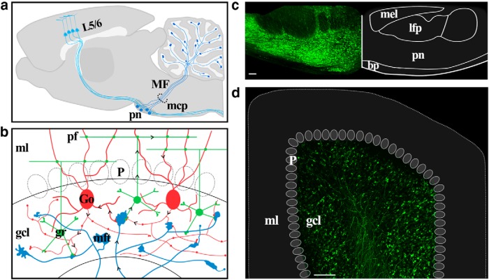
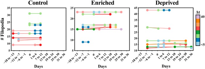
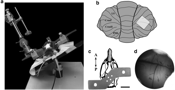
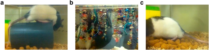
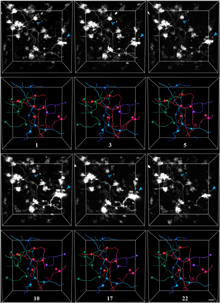
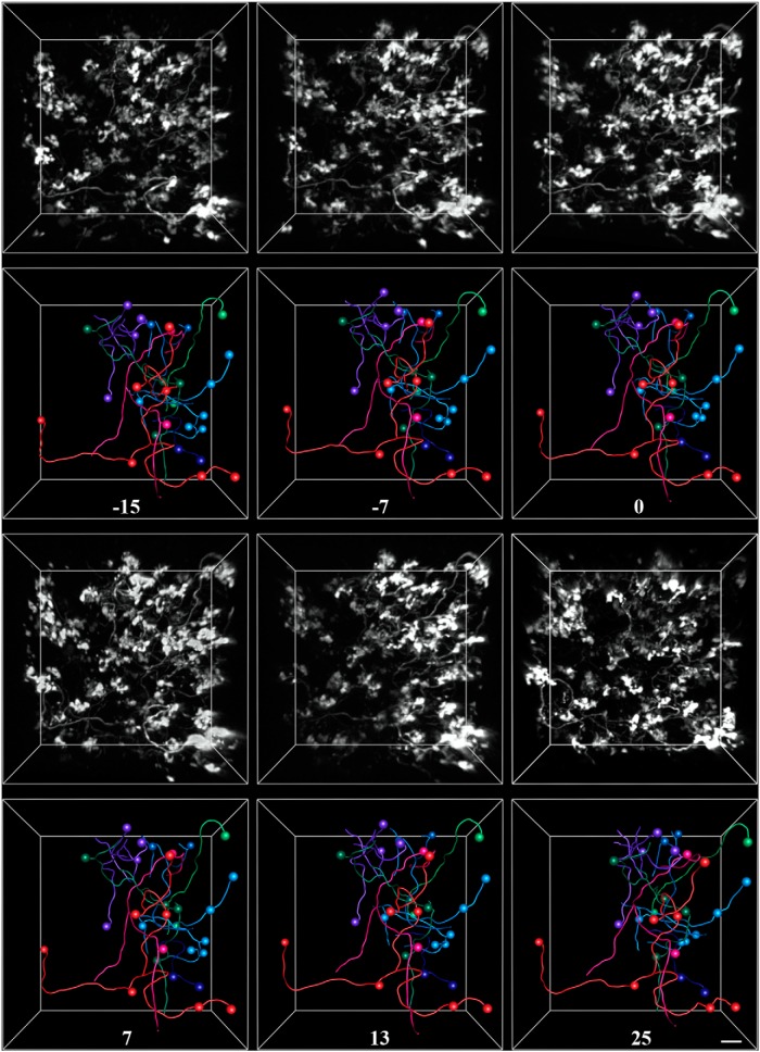
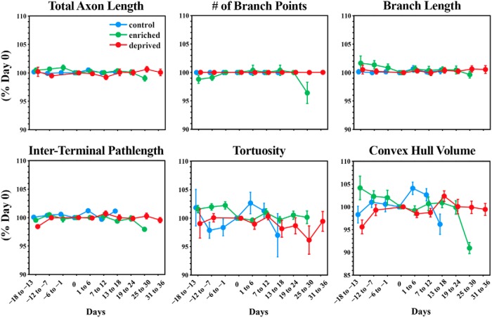
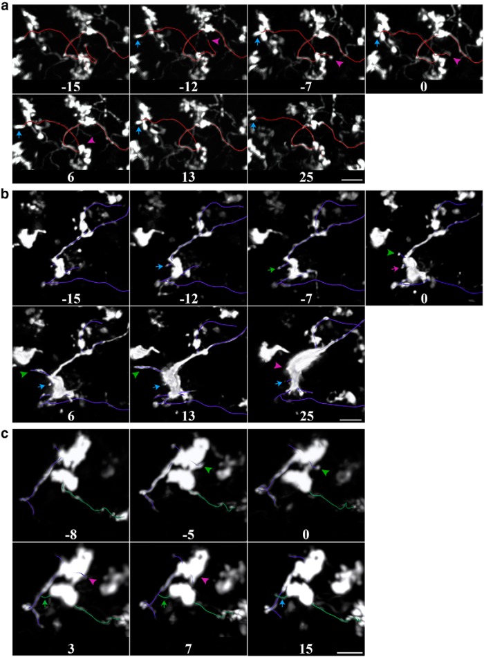
The cerebellum receives extensive disynaptic input from the neocortex via the basal pontine nuclei, the neurons of which send mossy fiber (MF) axons to the granule cell layer of the contralateral cerebellar hemisphere. Although this cortico-cerebellar circuit has been implicated in tasks such as sensory discrimination and motor learning, little is known about the potential role of MF morphological plasticity in the function of the cerebellar granule cell layer. To address this issue, we labeled MFs with EGFP via viral infection of the basal pons in adult rats and performed in vivo two-photon imaging of MFs in Crus I/II of the cerebellar hemisphere over a period of several weeks. Following the acquisition of baseline images, animals were housed in control, enriched, or deprived sensory environments. Morphological dynamics were assessed by tracing MF axons and their terminals, and by tracking the stability of filopodia arising from MF terminal rosettes. MF axons and terminals were found to be remarkably stable. Parameters derived neither from measurements of axonal arbor geometry nor from the morphology of individual rosettes and their filopodial extensions significantly changed under control conditions over 4 weeks of imaging. Increasing whisker stimulation by manipulating the sensory environment or decreasing such stimulation by whisker trimming also failed to alter MF structure. Our studies indicate that pontine MF axons projecting to Crus I/II in adult rats do not undergo significant structural rearrangements over the course of weeks, and that this stability is not altered by the sustained manipulation of whisker sensorimotor experience.
小脑通过基底脑桥核从新皮层接收广泛的双突触输入,基底脑桥核的神经元通过苔藓纤维(MF)轴突将其发送到对侧小脑半球的颗粒细胞层。尽管这个皮质-小脑回路已被认为参与了感觉辨别和运动学习等任务,但对于 MF 形态可塑性在小脑颗粒细胞层功能中的潜在作用知之甚少。为了解决这个问题,我们通过对成年大鼠基底脑桥的病毒感染来标记 MF,并在数周内对小脑半球 Crus I/II 中的 MF 进行体内双光子成像。在获得基线图像后,将动物饲养在对照、丰富或剥夺感觉环境中。通过追踪 MF 轴突及其末端,并通过跟踪 MF 末端玫瑰花结产生的稳定丝状伪足来评估形态动力学。MF 轴突和末端非常稳定。在 4 周的成像过程中,无论是从轴突分支几何形状的测量中还是从单个玫瑰花结及其丝状伪足的形态中得出的参数,都没有明显变化。通过操纵感觉环境增加胡须刺激或通过修剪胡须减少这种刺激也不能改变 MF 结构。我们的研究表明,成年大鼠投射到 Crus I/II 的脑桥 MF 轴突在数周内不会发生显著的结构重排,并且这种稳定性不会因持续操纵胡须感觉运动经验而改变。