• 文献检索
  • 文档翻译
  • 深度研究
  • 学术资讯
  • Suppr Zotero 插件Zotero 插件
  • 邀请有礼
  • 套餐&价格
  • 历史记录
应用&插件
Suppr Zotero 插件Zotero 插件浏览器插件Mac 客户端Windows 客户端微信小程序
定价
高级版会员购买积分包购买API积分包
服务
文献检索文档翻译深度研究API 文档MCP 服务
关于我们
关于 Suppr公司介绍联系我们用户协议隐私条款
关注我们

Suppr 超能文献

核心技术专利:CN118964589B侵权必究
粤ICP备2023148730 号-1Suppr @ 2026

文献检索

告别复杂PubMed语法,用中文像聊天一样搜索,搜遍4000万医学文献。AI智能推荐,让科研检索更轻松。

立即免费搜索

文件翻译

保留排版,准确专业,支持PDF/Word/PPT等文件格式,支持 12+语言互译。

免费翻译文档

深度研究

AI帮你快速写综述,25分钟生成高质量综述,智能提取关键信息,辅助科研写作。

立即免费体验

基于重建人表皮-Episkin®的红色毛癣菌感染模型

A Trichophyton Rubrum Infection Model Based on the Reconstructed Human Epidermis - Episkin®.

作者信息

Liang Pan-Pan, Huang Xin-Zhu, Yi Jin-Ling, Chen Zhi-Rui, Ma Han, Ye Cong-Xiu, Chen Xian-Yan, Lai Wei, Chen Jian

机构信息

Department of Dermatology, Third Affiliated Hospital of Sun Yat-sen University, Guangzhou, Guangdong 510630, China.

出版信息

Chin Med J (Engl). 2016 Jan 5;129(1):54-8. doi: 10.4103/0366-6999.172573.

DOI:10.4103/0366-6999.172573
PMID:26712433
原文链接:https://pmc.ncbi.nlm.nih.gov/articles/PMC4797543/
Abstract

BACKGROUND

Trichophyton rubrum represents the most common infectious fungus responsible for dermatophytosis in human, but the mechanism involved is still not completely understood. An appropriate model constructed to simulate host infection is the prerequisite to study the pathogenesis of dermatophytosis caused by T. rubrum. In this study, we intended to develop a new T. rubrum infection model in vitro, using the three-dimensional reconstructed epidermis - EpiSkin ®, and to pave the way for further investigation of the mechanisms involved in T. rubrum infection.

METHODS

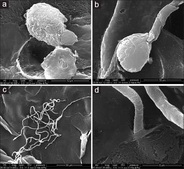
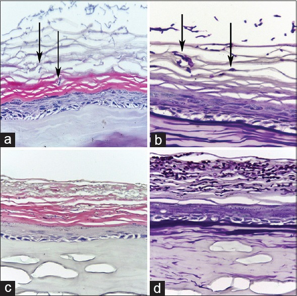
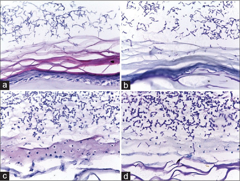
The reconstructed human epidermis (RHE) was infected by inoculating low-dose (400 conidia) and high-dose (4000 conidia) T. rubrum conidia to optimize the infection dose. During the various periods after infection, the samples were processed for pathological examination and scanning electron microscopy (SEM) observation.

RESULTS

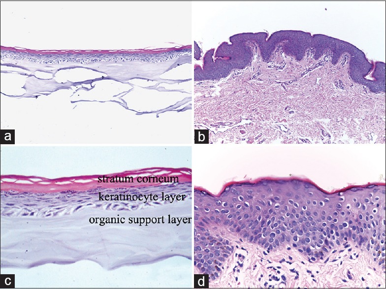
The histological analysis of RHE revealed a fully differentiated epidermis with a functional stratum corneum, which was analogous to the normal human epidermis. The results of hematoxylin and eosin staining and the periodic acid-Schiff staining showed that the infection dose of 400 conidia was in accord with the pathological characteristics of host dermatophytosis caused by T. rubrum. SEM observations further exhibited the process of T. rubrum infection in an intuitionistic way.

CONCLUSIONS

We established the T. rubrum infection model on RHE in vitro successfully. It is a promising model for further investigation of the mechanisms involved in T. rubrum infection.

摘要

背景

红色毛癣菌是引起人类皮肤癣菌病最常见的感染性真菌,但其致病机制仍未完全明确。构建合适的模拟宿主感染的模型是研究红色毛癣菌所致皮肤癣菌病发病机制的前提。在本研究中,我们旨在利用三维重建表皮-EpiSkin®建立一种新的体外红色毛癣菌感染模型,为进一步研究红色毛癣菌感染的机制铺平道路。

方法

通过接种低剂量(400个分生孢子)和高剂量(4000个分生孢子)的红色毛癣菌分生孢子感染重建人表皮(RHE),以优化感染剂量。在感染后的不同时期,对样本进行病理检查和扫描电子显微镜(SEM)观察。

结果

RHE的组织学分析显示其表皮完全分化,角质层功能正常,类似于正常人类表皮。苏木精-伊红染色和过碘酸-希夫染色结果表明,400个分生孢子的感染剂量符合红色毛癣菌引起的宿主皮肤癣菌病的病理特征。SEM观察进一步直观地展示了红色毛癣菌的感染过程。

结论

我们成功地在体外RHE上建立了红色毛癣菌感染模型。它是进一步研究红色毛癣菌感染机制的一个有前景的模型。

https://cdn.ncbi.nlm.nih.gov/pmc/blobs/4ccd/4797543/028d546bdfdf/CMJ-129-54-g004.jpg
https://cdn.ncbi.nlm.nih.gov/pmc/blobs/4ccd/4797543/cd8646a98384/CMJ-129-54-g001.jpg
https://cdn.ncbi.nlm.nih.gov/pmc/blobs/4ccd/4797543/0ba662b61cd8/CMJ-129-54-g002.jpg
https://cdn.ncbi.nlm.nih.gov/pmc/blobs/4ccd/4797543/a44fb5d0a85c/CMJ-129-54-g003.jpg
https://cdn.ncbi.nlm.nih.gov/pmc/blobs/4ccd/4797543/028d546bdfdf/CMJ-129-54-g004.jpg

相似文献

1
A Trichophyton Rubrum Infection Model Based on the Reconstructed Human Epidermis - Episkin®.基于重建人表皮-Episkin®的红色毛癣菌感染模型
Chin Med J (Engl). 2016 Jan 5;129(1):54-8. doi: 10.4103/0366-6999.172573.
2
Modeling dermatophytosis in reconstructed human epidermis: A new tool to study infection mechanisms and to test antifungal agents.在重建人表皮中模拟皮肤癣菌病:一种研究感染机制和测试抗真菌药物的新工具。
Med Mycol. 2017 Jul 1;55(5):485-494. doi: 10.1093/mmy/myw111.
3
Transcription profile of Trichophyton rubrum conidia grown on keratin reveals the induction of an adhesin-like protein gene with a tandem repeat pattern.在角蛋白上生长的红色毛癣菌分生孢子的转录谱揭示了一个具有串联重复模式的粘附素样蛋白基因的诱导。
BMC Genomics. 2016 Mar 18;17:249. doi: 10.1186/s12864-016-2567-8.
4
Responses of Reconstructed Human Epidermis to Trichophyton rubrum Infection and Impairment of Infection by the Inhibitor PD169316.重建人类表皮对红色毛癣菌感染的反应和抑制剂 PD169316 对感染的抑制作用。
J Invest Dermatol. 2019 Oct;139(10):2080-2089.e6. doi: 10.1016/j.jid.2019.03.1147. Epub 2019 Apr 12.
5
Evaluation of an Explanted Porcine Skin Model to Investigate Infection with the Dermatophyte Trichophyton rubrum.评价一种用于研究皮肤癣菌红色毛癣菌感染的猪皮模型。
Mycopathologia. 2020 Apr;185(2):233-243. doi: 10.1007/s11046-020-00438-9. Epub 2020 Feb 27.
6
An ultrastructural study of Trichophyton rubrum induced onychomycosis.红色毛癣菌所致甲真菌病的超微结构研究。
BMC Infect Dis. 2015 Nov 17;15:532. doi: 10.1186/s12879-015-1240-1.
7
Chronic dermatophytosis: what is special about Trichophyton rubrum?慢性皮肤癣菌病:红色毛癣菌有何特殊之处?
Adv Dermatol. 1994;9:97-109; discussion 110-1.
8
Trichophyton rubrum conidia modulate the expression and transport of Toll-like receptor 2 in HaCaT cell.红色毛癣菌分生孢子调节HaCaT细胞中Toll样受体2的表达和转运。
Microb Pathog. 2015 Jun-Jul;83-84:1-5. doi: 10.1016/j.micpath.2015.04.002. Epub 2015 Apr 11.
9
[Establishing an experimental guinea pig model of dermatophytosis Using Trichophyton rubrum].[利用红色毛癣菌建立豚鼠皮肤癣菌病实验模型]
Zhongguo Yi Xue Ke Xue Yuan Xue Bao. 2008 Oct;30(5):599-602.
10
In vitro and ex vivo infection models help assess the molecular aspects of the interaction of Trichophyton rubrum with the host milieu.体外和离体感染模型有助于评估红色毛癣菌与宿主环境相互作用的分子方面。
Med Mycol. 2016 May;54(4):420-7. doi: 10.1093/mmy/myv113. Epub 2016 Jan 14.

引用本文的文献

1
In vitro characterization of Trichophyton rubrum biofilm by combined anti-biofilm enzymes.联合抗生物膜酶对红色毛癣菌生物膜的体外特性研究
PLoS One. 2025 Sep 10;20(9):e0331291. doi: 10.1371/journal.pone.0331291. eCollection 2025.
2
Innate immune responses against the fungal pathogen Candida auris.固有免疫应答针对真菌病原体耳念珠菌。
Nat Commun. 2022 Jun 21;13(1):3553. doi: 10.1038/s41467-022-31201-x.
3
Towards a Standardized Procedure for the Production of Infective Spores to Study the Pathogenesis of Dermatophytosis.

本文引用的文献

1
Establishment and characterization of a reconstructed Chinese human epidermis model.一种重建的中国人表皮模型的建立与表征
Int J Cosmet Sci. 2016 Feb;38(1):60-7. doi: 10.1111/ics.12249. Epub 2015 Aug 24.
2
Trichophyton rubrum conidia modulate the expression and transport of Toll-like receptor 2 in HaCaT cell.红色毛癣菌分生孢子调节HaCaT细胞中Toll样受体2的表达和转运。
Microb Pathog. 2015 Jun-Jul;83-84:1-5. doi: 10.1016/j.micpath.2015.04.002. Epub 2015 Apr 11.
3
Dermatophytes activate skin keratinocytes via mitogen-activated protein kinase signaling and induce immune responses.
迈向生产感染性孢子的标准化程序以研究皮肤癣菌病的发病机制
J Fungi (Basel). 2021 Nov 30;7(12):1029. doi: 10.3390/jof7121029.
4
Challenges in exploring and manipulating the human skin microbiome.探索和操纵人类皮肤微生物组的挑战。
Microbiome. 2021 May 30;9(1):125. doi: 10.1186/s40168-021-01062-5.
5
Human skin models: From healthy to disease-mimetic systems; characteristics and applications.人体皮肤模型:从健康到疾病模拟系统;特点与应用。
Br J Pharmacol. 2020 Oct;177(19):4314-4329. doi: 10.1111/bph.15184. Epub 2020 Aug 19.
6
Corrigendum: A Trichophyton Rubrum infection model based on the reconstructed human epidermis - Episkin®.勘误:基于重建人表皮-Episkin®的红色毛癣菌感染模型。
Chin Med J (Engl). 2019 Sep 20;132(18):2268. doi: 10.1097/CM9.0000000000000452.
7
An Human Skin Model to Study Superficial Fungal Infections.一种用于研究浅表真菌感染的人体皮肤模型。
Front Microbiol. 2019 Jun 5;10:1172. doi: 10.3389/fmicb.2019.01172. eCollection 2019.
皮肤癣菌通过丝裂原活化蛋白激酶信号传导激活皮肤角质形成细胞并诱导免疫反应。
Infect Immun. 2015 Apr;83(4):1705-14. doi: 10.1128/IAI.02776-14. Epub 2015 Feb 9.
4
Infection of keratinocytes with Trichophytum rubrum induces epidermal growth factor-dependent RNase 7 and human beta-defensin-3 expression.红色毛癣菌感染角质形成细胞可诱导表皮生长因子依赖性核糖核酸酶7和人β-防御素-3的表达。
PLoS One. 2014 Apr 18;9(4):e93941. doi: 10.1371/journal.pone.0093941. eCollection 2014.
5
Mycology - an update. Part 1: Dermatomycoses: causative agents, epidemiology and pathogenesis.真菌学——最新进展。第1部分:皮肤真菌病:病原体、流行病学及发病机制。
J Dtsch Dermatol Ges. 2014 Mar;12(3):188-209; quiz 210, 188-211; quiz 212. doi: 10.1111/ddg.12245. Epub 2014 Feb 17.
6
Extensive deep dermatophytosis cause by Trichophyton rubrum in a patient with liver cirrhosis and chronic renal failure.广泛深部皮肤癣菌病由红色毛癣菌引起,患者患有肝硬化和慢性肾衰竭。
Mycopathologia. 2013 Dec;176(5-6):457-62. doi: 10.1007/s11046-013-9696-2. Epub 2013 Aug 28.
7
Exposure to heat-inactivated Trichophyton rubrum resulting in a limited immune response of human keratinocytes.暴露于热灭活红色毛癣菌导致人类角质形成细胞的有限免疫反应。
Chin Med J (Engl). 2013 Jan;126(2):215-9.
8
Deep Trichophyton rubrum infection presenting with umbilicated papulonodules in a cardiac transplant recipient.一名心脏移植受者出现脐凹性丘疹结节的红色毛癣菌深部感染。
Mycoses. 2013 May;56(3):361-4. doi: 10.1111/myc.12001. Epub 2012 Sep 14.
9
Animal model of dermatophytosis.皮肤癣菌病动物模型
J Biomed Biotechnol. 2012;2012:125384. doi: 10.1155/2012/125384. Epub 2012 Apr 29.
10
Mechanisms of skin adherence and invasion by dermatophytes.皮肤癣菌的皮肤黏附与侵袭机制。
Mycoses. 2012 May;55(3):218-23. doi: 10.1111/j.1439-0507.2011.02081.x. Epub 2011 Aug 11.