Yue Monica S, Plavicki Jessica S, Li Xin-yi, Peterson Richard E, Heideman Warren
Molecular and Environmental Toxicology Center, University of Wisconsin, 1300 University Avenue, Madison, WI, 53706, USA.
Pharmaceutical Sciences Division, School of Pharmacy, University of Wisconsin, 777 Highland Avenue, Madison, WI, 53705, USA.
BMC Dev Biol. 2015 Dec 29;15:50. doi: 10.1186/s12861-015-0100-y.
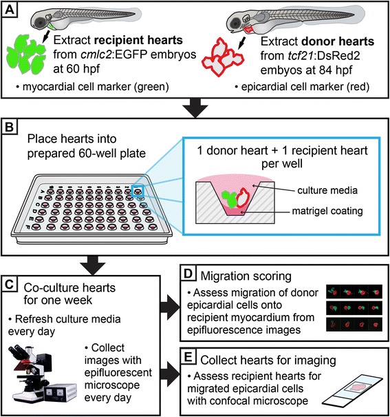
The vertebrate heart consists of three cell layers: the innermost endothelium, the contractile myocardium and the outermost epicardium. The epicardium is vital for heart development and function, and forms from epicardial progenitor cells (EPCs), which migrate to the myocardium during early development. Disruptions in EPC migration and epicardium formation result in a number of cardiac malformations, many of which resemble congenital heart diseases in humans. Hence, it is important to understand the mechanisms that influence EPC migration and spreading in the developing heart. In vitro approaches heretofore have been limited to monolayer epicardial cell cultures, which may not fully capture the complex interactions that can occur between epicardial and myocardial cells in vivo.
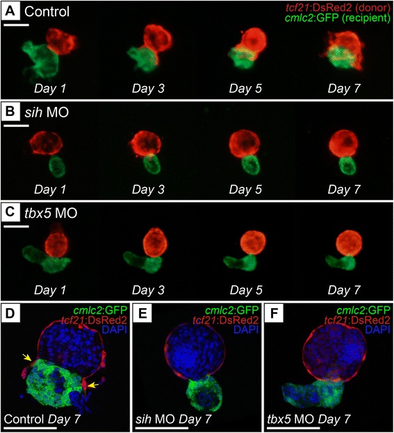
Here we describe a novel in vitro co-culture assay for assessing epicardial cell migration using embryonic zebrafish hearts. We isolated donor hearts from embryonic zebrafish carrying an epicardial-specific fluorescent reporter after epicardial cells were present on the heart. These were co-cultured with recipient hearts expressing a myocardial-specific fluorescent reporter, isolated prior to EPC migration. Using this method, we can clearly visualize the movement of epicardial cells from the donor heart onto the myocardium of the recipient heart. We demonstrate the utility of this method by showing that epicardial cell migration is significantly delayed or absent when myocardial cells lack contractility and when myocardial cells are deficient in tbx5 expression.
We present a method to assess the migration of epicardial cells in an in vitro assay, wherein the migration of epicardial cells from a donor heart onto the myocardium of a recipient heart in co-culture is monitored and scored. The donor and recipient hearts can be independently manipulated, using either genetic tools or pharmacological agents. This allows flexibility in experimental design for determining the role that target genes/signaling pathways in specific cell types may have on epicardial cell migration.
脊椎动物的心脏由三层细胞组成:最内层的内皮、收缩性的心肌和最外层的心外膜。心外膜对心脏发育和功能至关重要,由心外膜祖细胞(EPCs)形成,这些祖细胞在早期发育过程中迁移至心肌。EPC迁移和心外膜形成的破坏会导致多种心脏畸形,其中许多类似于人类的先天性心脏病。因此,了解影响发育中心脏中EPC迁移和扩散的机制很重要。迄今为止,体外方法仅限于单层心外膜细胞培养,这可能无法完全捕捉体内心外膜和心肌细胞之间可能发生的复杂相互作用。
在此,我们描述了一种使用胚胎斑马鱼心脏评估心外膜细胞迁移的新型体外共培养测定法。在心脏上出现心外膜细胞后,我们从携带心外膜特异性荧光报告基因的胚胎斑马鱼中分离出供体心脏。将这些供体心脏与在EPC迁移之前分离的、表达心肌特异性荧光报告基因的受体心脏进行共培养。使用这种方法,我们可以清楚地观察到心外膜细胞从供体心脏移动到受体心脏的心肌上。我们通过显示当心肌细胞缺乏收缩力以及当心肌细胞中tbx5表达不足时心外膜细胞迁移显著延迟或缺失,证明了该方法的实用性。
我们提出了一种在体外测定中评估心外膜细胞迁移的方法,其中监测并记录共培养时心外膜细胞从供体心脏迁移到受体心脏心肌上的情况。供体和受体心脏可以使用基因工具或药物进行独立操作。这为确定特定细胞类型中的靶基因/信号通路可能对心外膜细胞迁移所起的作用提供了实验设计的灵活性。