Liao Naishun, Wu Ming, Pan Fan, Lin Jiumao, Li Zuanfang, Zhang Da, Wang Yingchao, Zheng Youshi, Peng Jun, Liu Xiaolong, Liu Jingfeng
The United Innovation of Mengchao Hepatobiliary Technology Key Laboratory of Fujian Province, Mengchao Hepatobiliary Hospital of Fujian Medical University, Fuzhou 350025, P.R. China.
The Liver Center of Fujian Province, Fujian Medical University, Fuzhou 350025, P.R. China.
Sci Rep. 2016 Jan 5;6:18746. doi: 10.1038/srep18746.
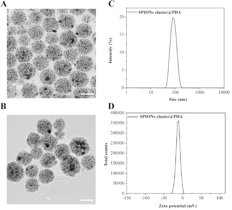
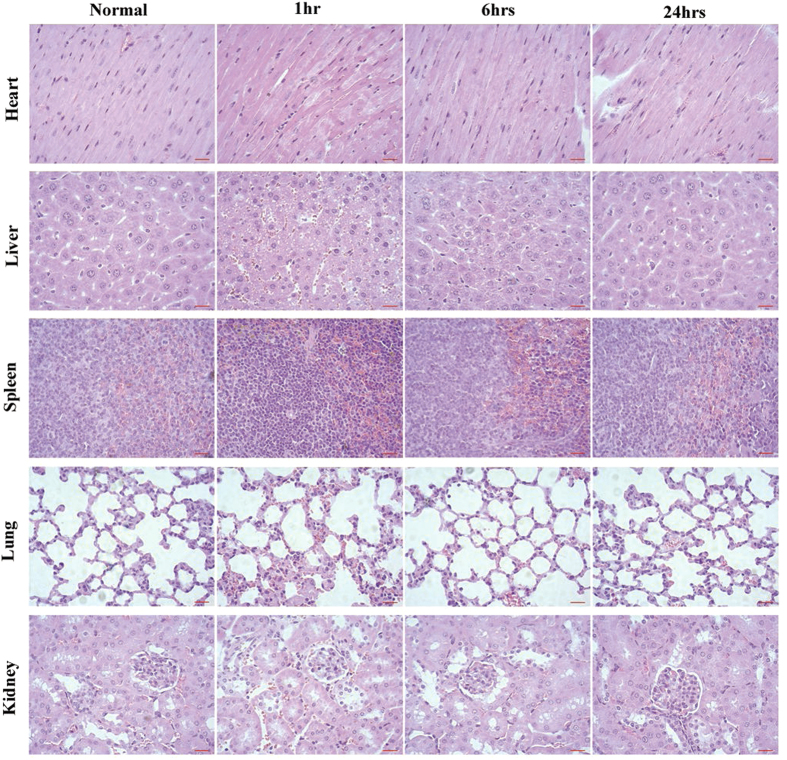
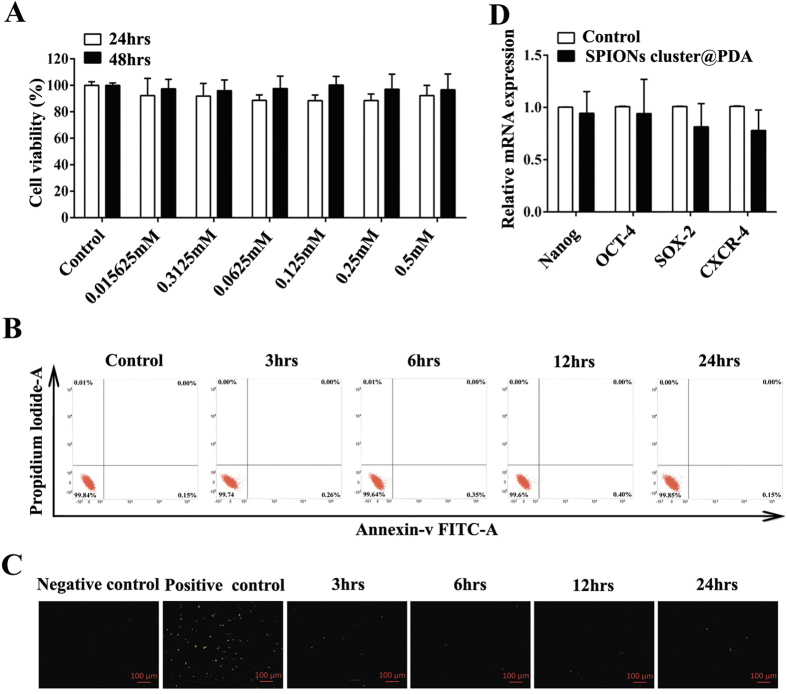
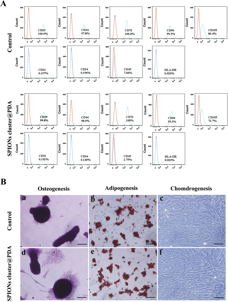
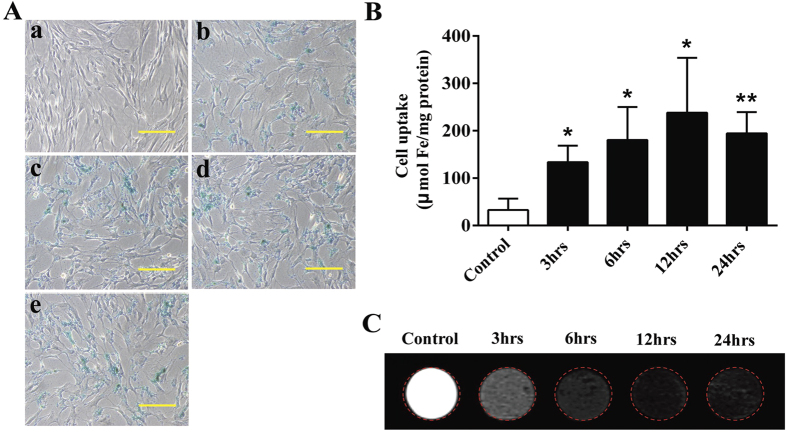
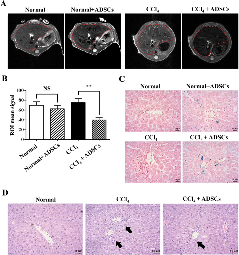
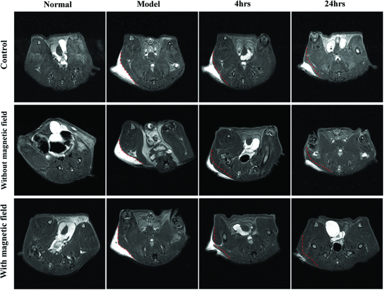
Tracking and monitoring of cells in vivo after transplantation can provide crucial information for stem cell therapy. Magnetic resonance imaging (MRI) combined with contrast agents is believed to be an effective and non-invasive technique for cell tracking in living bodies. However, commercial superparamagnetic iron oxide nanoparticles (SPIONs) applied to label cells suffer from shortages such as potential toxicity, low labeling efficiency, and low contrast enhancing. Herein, the adipose tissue-derived stem cells (ADSCs) were efficiently labeled with SPIONs coated with poly (dopamine) (SPIONs cluster@PDA), without affecting their viability, proliferation, apoptosis, surface marker expression, as well as their self-renew ability and multi-differentiation potential. The labeled cells transplanted into the mice through tail intravenous injection exhibited a negative enhancement of the MRI signal in the damaged liver-induced by carbon tetrachloride, and subsequently these homed ADSCs with SPIONs cluster@PDA labeling exhibited excellent repair effects to the damaged liver. Moreover, the enhanced target-homing to tissue of interest and repair effects of SPIONs cluster@PDA-labeled ADSCs could be achieved by use of external magnetic field in the excisional skin wound mice model. Therefore, we provide a facile, safe, noninvasive and sensitive method for external magnetic field targeted delivery and MRI based tracking of transplanted cells in vivo.
移植后对体内细胞进行追踪和监测可为干细胞治疗提供关键信息。磁共振成像(MRI)结合造影剂被认为是一种用于活体细胞追踪的有效且非侵入性的技术。然而,用于标记细胞的商业超顺磁性氧化铁纳米颗粒(SPIONs)存在诸如潜在毒性、低标记效率和低对比度增强等缺点。在此,脂肪组织来源的干细胞(ADSCs)被聚(多巴胺)包覆的SPIONs(SPIONs簇@PDA)有效标记,且不影响其活力、增殖、凋亡、表面标志物表达以及自我更新能力和多向分化潜能。通过尾静脉注射将标记的细胞移植到小鼠体内后,在四氯化碳诱导的受损肝脏中显示出MRI信号的负增强,随后这些带有SPIONs簇@PDA标记的归巢ADSCs对受损肝脏表现出优异的修复效果。此外,在切除性皮肤伤口小鼠模型中,通过使用外部磁场可实现SPIONs簇@PDA标记的ADSCs对感兴趣组织的增强靶向归巢和修复效果。因此,我们提供了一种简便、安全、非侵入性且灵敏的方法,用于在体内对移植细胞进行外部磁场靶向递送和基于MRI的追踪。