Sleigh Alison, Savage David B, Williams Guy B, Porter David, Carpenter T Adrian, Brindle Kevin M, Kemp Graham J
Wolfson Brain Imaging Centre, University of Cambridge School of Clinical Medicine, Cambridge Biomedical Campus, United Kingdom; National Institute for Health Research/Wellcome Trust Clinical Research Facility at Cambridge University Hospitals NHS Foundation Trust, Cambridge Biomedical Campus, United Kingdom;
University of Cambridge Metabolic Research Laboratories, Wellcome Trust-Medical Research Council Institute of Metabolic Science, Cambridge Biomedical Campus, United Kingdom;
J Appl Physiol (1985). 2016 Mar 15;120(6):649-56. doi: 10.1152/japplphysiol.00871.2015. Epub 2016 Jan 7.
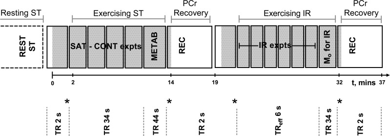
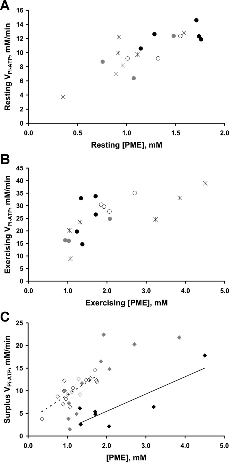
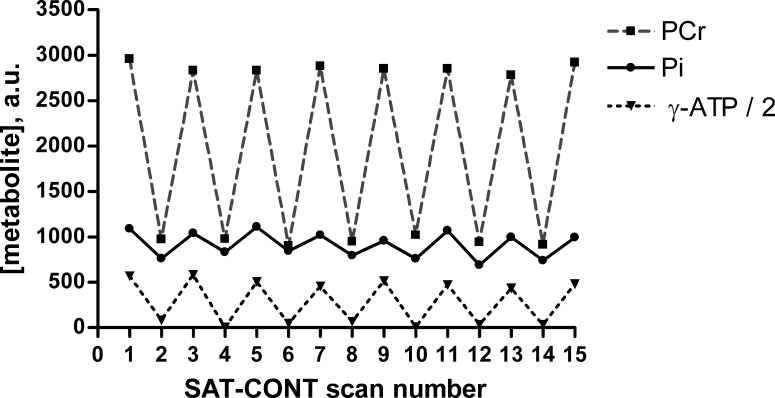
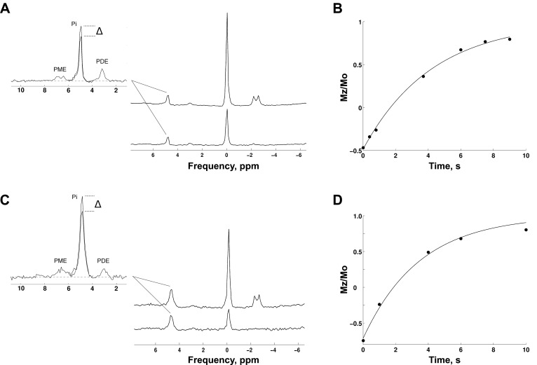
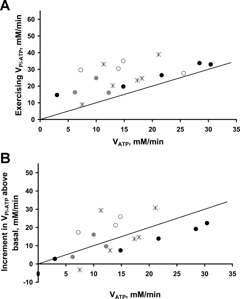
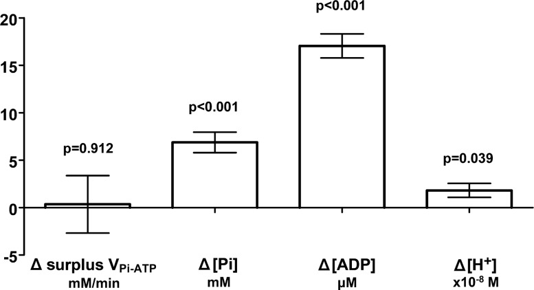
Fundamental criticisms have been made over the use of (31)P magnetic resonance spectroscopy (MRS) magnetization transfer estimates of inorganic phosphate (Pi)→ATP flux (VPi-ATP) in human resting skeletal muscle for assessing mitochondrial function. Although the discrepancy in the magnitude of VPi-ATP is now acknowledged, little is known about its metabolic determinants. Here we use a novel protocol to measure VPi-ATP in human exercising muscle for the first time. Steady-state VPi-ATP was measured at rest and over a range of exercise intensities and compared with suprabasal oxidative ATP synthesis rates estimated from the initial rates of postexercise phosphocreatine resynthesis (VATP). We define a surplus Pi→ATP flux as the difference between VPi-ATP and VATP. The coupled reactions catalyzed by the glycolytic enzymes GAPDH and phosphoglycerate kinase (PGK) have been shown to catalyze measurable exchange between ATP and Pi in some systems and have been suggested to be responsible for this surplus flux. Surplus VPi-ATP did not change between rest and exercise, even though the concentrations of Pi and ADP, which are substrates for GAPDH and PGK, respectively, increased as expected. However, involvement of these enzymes is suggested by correlations between absolute and surplus Pi→ATP flux, both at rest and during exercise, and the intensity of the phosphomonoester peak in the (31)P NMR spectrum. This peak includes contributions from sugar phosphates in the glycolytic pathway, and changes in its intensity may indicate changes in downstream glycolytic intermediates, including 3-phosphoglycerate, which has been shown to influence the exchange between ATP and Pi catalyzed by GAPDH and PGK.
对于在人体静息骨骼肌中使用(31)P磁共振波谱(MRS)磁化转移估计无机磷酸盐(Pi)→ATP通量(VPi-ATP)来评估线粒体功能,已经有人提出了根本性的批评。尽管现在已经认识到VPi-ATP大小存在差异,但其代谢决定因素却知之甚少。在这里,我们首次使用一种新方案来测量人体运动肌肉中的VPi-ATP。在静息状态和一系列运动强度下测量稳态VPi-ATP,并与根据运动后磷酸肌酸再合成的初始速率估计的超基础氧化ATP合成速率(VATP)进行比较。我们将多余的Pi→ATP通量定义为VPi-ATP与VATP之间的差值。糖酵解酶甘油醛-3-磷酸脱氢酶(GAPDH)和磷酸甘油酸激酶(PGK)催化的偶联反应已被证明在某些系统中能催化ATP和Pi之间可测量的交换,并被认为是造成这种多余通量的原因。尽管分别作为GAPDH和PGK底物的Pi和ADP浓度如预期那样增加,但静息和运动之间多余的VPi-ATP并没有变化。然而,在静息和运动期间,绝对和多余的Pi→ATP通量与(31)P NMR谱中磷酸单酯峰的强度之间的相关性表明了这些酶的参与。这个峰包括糖酵解途径中糖磷酸的贡献,其强度的变化可能表明下游糖酵解中间产物的变化,包括3-磷酸甘油酸,已证明它会影响GAPDH和PGK催化的ATP与Pi之间的交换。