• 文献检索
  • 文档翻译
  • 深度研究
  • 学术资讯
  • Suppr Zotero 插件Zotero 插件
  • 邀请有礼
  • 套餐&价格
  • 历史记录
应用&插件
Suppr Zotero 插件Zotero 插件浏览器插件Mac 客户端Windows 客户端微信小程序
定价
高级版会员购买积分包购买API积分包
服务
文献检索文档翻译深度研究API 文档MCP 服务
关于我们
关于 Suppr公司介绍联系我们用户协议隐私条款
关注我们

Suppr 超能文献

核心技术专利:CN118964589B侵权必究
粤ICP备2023148730 号-1Suppr @ 2026

文献检索

告别复杂PubMed语法,用中文像聊天一样搜索,搜遍4000万医学文献。AI智能推荐,让科研检索更轻松。

立即免费搜索

文件翻译

保留排版,准确专业,支持PDF/Word/PPT等文件格式,支持 12+语言互译。

免费翻译文档

深度研究

AI帮你快速写综述,25分钟生成高质量综述,智能提取关键信息,辅助科研写作。

立即免费体验

大鼠致密骨的哈弗斯系统及使用三维重建对骨内膜侧和骨膜侧的比较

Haversian system of compact bone and comparison between endosteal and periosteal sides using three-dimensional reconstruction in rat.

作者信息

Kim Jeong-Nam, Lee Jun-Young, Shin Kang-Jae, Gil Young-Chul, Koh Ki-Seok, Song Wu-Chul

机构信息

Department of Biomedical Laboratory Science, Masan University, Masan, Korea.

Department of Anatomy, Research Institute of Medical Science, Konkuk University School of Medicine, Seoul, Korea.

出版信息

Anat Cell Biol. 2015 Dec;48(4):258-61. doi: 10.5115/acb.2015.48.4.258. Epub 2015 Dec 21.

DOI:10.5115/acb.2015.48.4.258
PMID:26770876
原文链接:https://pmc.ncbi.nlm.nih.gov/articles/PMC4701699/
Abstract

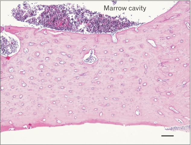
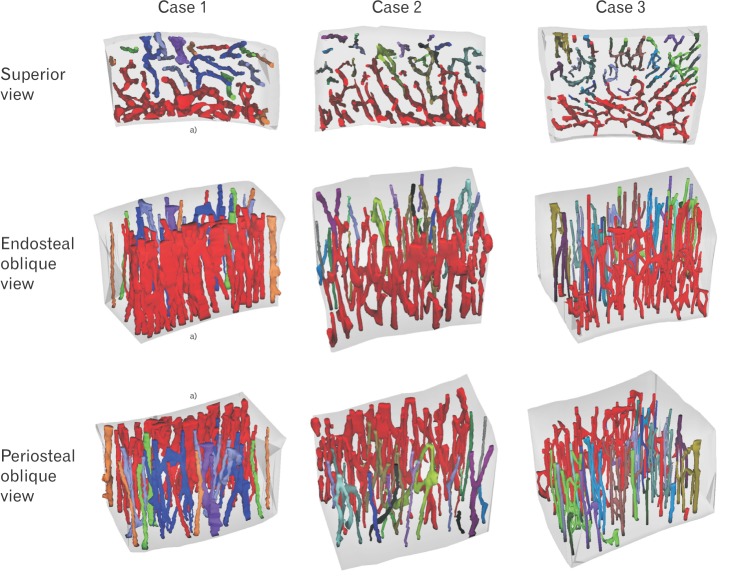
The current model of compact bone is that of a system of Haversian (longitudinal) canals connected by Volkmann's (transverse) canals. Models based on either histology or microcomputed tomography do not accurately represent the morphologic detail and microstructure of this system, especially that of the canal networks and their spatial relationships. The aim of the present study was to demonstrate the morphologic pattern and network of the Haversian system and to compare endosteal and periosteal sides in rats using three-dimensional (3D) reconstruction. Ten Sprague-Dawley rats aged 8-10 weeks were used. The femurs were harvested from each rat and fixed, decalcified with 10% EDTA-2Na, serially sectioned at a thickness of 5 µm, and then stained with hematoxylin and eosin. The serial sections were reconstructed three-dimensionally using Reconstruct software. The Haversian canals in the endosteal region were found to be large, highly interconnected, irregular, and close to neighboring canals. In contrast, the canals in the periosteal region were straight and small. This combined application of 3D reconstruction and histology examinations to the Haversian system has confirmed its microstructure, showing a branched network pattern on the endosteal side but not on the periosteal side.

摘要

目前密质骨的模型是由哈弗斯(纵向)管系统通过福尔克曼(横向)管相连组成的。基于组织学或显微计算机断层扫描的模型不能准确呈现该系统的形态细节和微观结构,尤其是管网络及其空间关系的细节。本研究的目的是利用三维(3D)重建技术展示大鼠哈弗斯系统的形态模式和网络,并比较骨内膜和骨膜侧的情况。选用了10只8 - 10周龄的斯普拉格 - 道利大鼠。从每只大鼠身上取下股骨并进行固定,用10%乙二胺四乙酸二钠(EDTA - 2Na)脱钙,以5微米的厚度连续切片,然后用苏木精和伊红染色。使用Reconstruct软件对连续切片进行三维重建。发现骨内膜区域的哈弗斯管较大、高度相互连接、不规则且靠近相邻的管道。相比之下,骨膜区域的管道则是直的且较小。这种将3D重建和组织学检查联合应用于哈弗斯系统的方法证实了其微观结构,显示骨内膜侧呈现分支网络模式,而骨膜侧则没有。

https://cdn.ncbi.nlm.nih.gov/pmc/blobs/30f6/4701699/a8fac322d8e0/acb-48-258-g002.jpg
https://cdn.ncbi.nlm.nih.gov/pmc/blobs/30f6/4701699/561bafebeaea/acb-48-258-g001.jpg
https://cdn.ncbi.nlm.nih.gov/pmc/blobs/30f6/4701699/a8fac322d8e0/acb-48-258-g002.jpg
https://cdn.ncbi.nlm.nih.gov/pmc/blobs/30f6/4701699/561bafebeaea/acb-48-258-g001.jpg
https://cdn.ncbi.nlm.nih.gov/pmc/blobs/30f6/4701699/a8fac322d8e0/acb-48-258-g002.jpg

相似文献

1
Haversian system of compact bone and comparison between endosteal and periosteal sides using three-dimensional reconstruction in rat.大鼠致密骨的哈弗斯系统及使用三维重建对骨内膜侧和骨膜侧的比较
Anat Cell Biol. 2015 Dec;48(4):258-61. doi: 10.5115/acb.2015.48.4.258. Epub 2015 Dec 21.
2
Anatomy of the intracortical canal system: scanning electron microscopy study in rabbit femur.皮质内管系统的解剖结构:兔股骨的扫描电子显微镜研究
Clin Orthop Relat Res. 2009 Sep;467(9):2446-56. doi: 10.1007/s11999-009-0806-x. Epub 2009 Mar 28.
3
A model of the intracortical vascular system of long bones and of its organization: an experimental study in rabbit femur and tibia.长骨皮质内脉管系统及其组织的模型:兔股骨和胫骨的实验研究。
J Anat. 2008 Aug;213(2):183-93. doi: 10.1111/j.1469-7580.2008.00932.x.
4
Three-dimensional microstructures of the intracortical canals in the animal model of osteoporosis.骨质疏松动物模型中皮质内微管的三维微观结构。
Anat Cell Biol. 2020 Jun 30;53(2):162-168. doi: 10.5115/acb.19.189.
5
Microradiographic and histomorphometric indices of mandibles for diagnosis of osteopenia.
Scand J Dent Res. 1982 Feb;90(1):47-63. doi: 10.1111/j.1600-0722.1982.tb01523.x.
6
[The system of bony canals as the basis for the angioarchitectonics of bones].[作为骨血管构筑学基础的骨管系统]
Arkh Anat Gistol Embriol. 1976 Apr;70(4):53-61.
7
Three-dimensional reconstruction of Haversian systems in human cortical bone using synchrotron radiation-based micro-CT: morphology and quantification of branching and transverse connections across age.利用基于同步辐射的显微CT对人类皮质骨哈弗斯系统进行三维重建:不同年龄阶段分支和横向连接的形态学及定量分析
J Anat. 2016 May;228(5):719-32. doi: 10.1111/joa.12430. Epub 2016 Jan 7.
8
[Significance of the haversian number].[哈弗斯管数量的意义]
Bull Assoc Anat (Nancy). 1978 Dec;62(179):411-7.
9
Characterization of rat vertebrae cortical bone microstructures using confocal Raman microscopy combined to tomography and electron microscopy.利用共聚焦拉曼显微镜结合断层扫描和电子显微镜对大鼠椎骨皮质骨微观结构进行表征。
Ann Anat. 2023 Oct;250:152162. doi: 10.1016/j.aanat.2023.152162. Epub 2023 Sep 27.
10
Vascular canals in bovine cortical bone studied by corrosion casting.通过腐蚀铸型法研究牛皮质骨中的血管通道。
Calcif Tissue Int. 1990 Nov;47(5):320-5. doi: 10.1007/BF02555916.

引用本文的文献

1
A review of osteoporotic vertebral fracture animal models.骨质疏松性椎体骨折动物模型综述。
Biomed Eng Online. 2025 Apr 7;24(1):40. doi: 10.1186/s12938-025-01372-x.
2
The Gross Anatomical and Histological Features of the Humerus in African Green Monkeys () from Saint Kitts and Nevis, West Indies.来自西印度群岛圣基茨和尼维斯的非洲绿猴()肱骨的大体解剖和组织学特征。
Life (Basel). 2024 Oct 12;14(10):1295. doi: 10.3390/life14101295.
3
Are Artificial Intelligence-Assisted Three-Dimensional Histological Reconstructions Reliable for the Assessment of Trabecular Microarchitecture?

本文引用的文献

1
A comparison between micro-CT and histology for the evaluation of cortical bone: effect of polymethylmethacrylate embedding on structural parameters.微计算机断层扫描与组织学评估皮质骨的比较:聚甲基丙烯酸甲酯包埋对结构参数的影响。
J Microsc. 2012 Mar;245(3):302-10. doi: 10.1111/j.1365-2818.2011.03573.x. Epub 2011 Nov 22.
2
Radiological and histologic studies of the mandibular cortex of ovariectomized monkeys.
Oral Surg Oral Med Oral Pathol Oral Radiol Endod. 2011 Mar;111(3):372-80. doi: 10.1016/j.tripleo.2010.12.003.
3
3D visualization and quantification of rat cortical bone porosity using a desktop micro-CT system: a case study in the tibia.使用桌面型微计算机断层扫描系统对大鼠皮质骨孔隙率进行 3D 可视化和定量分析:以胫骨为例的研究。
人工智能辅助的三维组织学重建对小梁微结构评估是否可靠?
J Clin Med. 2024 Feb 15;13(4):1106. doi: 10.3390/jcm13041106.
4
Factors Affecting Holmium Laser Efficiency: Comparison of Laryngeal Mask Airway and Endotracheal Intubation During Ureteroscopy for Renal Stones.影响钬激光效率的因素:输尿管镜取石术治疗肾结石时喉罩气道与气管插管的比较。
J Endourol. 2024 Jan;38(1):8-15. doi: 10.1089/end.2023.0294. Epub 2023 Dec 7.
5
Evaluation of Human Bone Marrow Mesenchymal Stromal Cell (MSC) Functions on a Biomorphic Rattan-Wood-Derived Scaffold: A Comparison between Cultured and Uncultured MSCs.人骨髓间充质基质细胞(MSC)在生物形态藤木衍生支架上的功能评估:培养的和未培养的MSC之间的比较
Bioengineering (Basel). 2021 Dec 21;9(1):1. doi: 10.3390/bioengineering9010001.
6
Bone Healing and Inflammation: Principles of Fracture and Repair.骨愈合与炎症:骨折与修复原理
Semin Plast Surg. 2021 Aug;35(3):198-203. doi: 10.1055/s-0041-1732334. Epub 2021 Sep 10.
7
Bone remodeling in the longest living rodent, the naked mole-rat: Interelement variation and the effects of reproduction.最长寿啮齿动物——裸鼹鼠的骨骼重塑:元素间的变化和繁殖的影响。
J Anat. 2021 Jul;239(1):81-100. doi: 10.1111/joa.13404. Epub 2021 Feb 7.
8
Osteon: Structure, Turnover, and Regeneration.骨单位:结构、转换和再生。
Tissue Eng Part B Rev. 2022 Apr;28(2):261-278. doi: 10.1089/ten.TEB.2020.0322. Epub 2021 Mar 8.
9
Three-dimensional microstructures of the intracortical canals in the animal model of osteoporosis.骨质疏松动物模型中皮质内微管的三维微观结构。
Anat Cell Biol. 2020 Jun 30;53(2):162-168. doi: 10.5115/acb.19.189.
10
Assessment of the Inner Surface Microstructure of Decellularized Cortical Bone by a Scanning Electron Microscope.利用扫描电子显微镜评估脱细胞皮质骨的内表面微观结构
Bioengineering (Basel). 2019 Sep 19;6(3):86. doi: 10.3390/bioengineering6030086.
J Microsc. 2010 Oct;240(1):32-7. doi: 10.1111/j.1365-2818.2010.03381.x.
4
Morphometric analysis of the canal system of cortical bone: An experimental study in the rabbit femur carried out with standard histology and micro-CT.皮质骨管系统的形态计量学分析:一项在兔股骨上进行的采用标准组织学和显微CT的实验研究。
Anat Histol Embryol. 2010 Feb;39(1):17-26. doi: 10.1111/j.1439-0264.2009.00973.x. Epub 2009 Oct 28.
5
Reconstruct: a free editor for serial section microscopy.Reconstruct:一款用于连续切片显微镜检查的免费编辑器。
J Microsc. 2005 Apr;218(Pt 1):52-61. doi: 10.1111/j.1365-2818.2005.01466.x.
6
Comparison of microcomputed tomographic and microradiographic measurements of cortical bone porosity.皮质骨孔隙度的显微计算机断层扫描和显微放射照相测量结果比较
Calcif Tissue Int. 2004 May;74(5):437-47. doi: 10.1007/s00223-003-0071-z. Epub 2004 Feb 17.