Department of Radiology, UT Southwestern Medical Center, 5323 Harry Hines Blvd., Dallas, TX 75390, USA.
Department of Mechanical Engineering, Southern Methodist University, Dallas, TX 75275, USA.
Diagnostics (Basel). 2013 Jul 9;3(3):325-43. doi: 10.3390/diagnostics3030325.
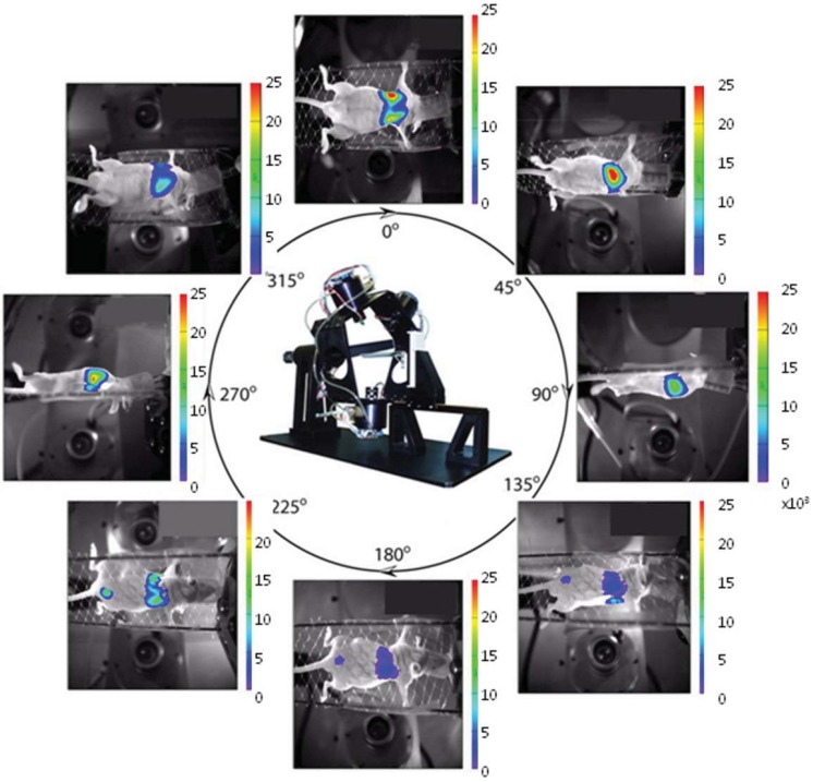
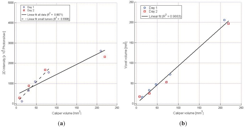
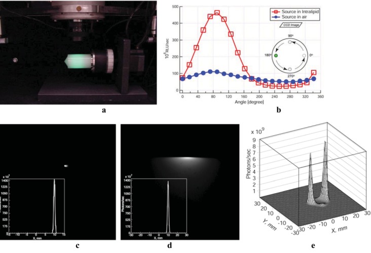
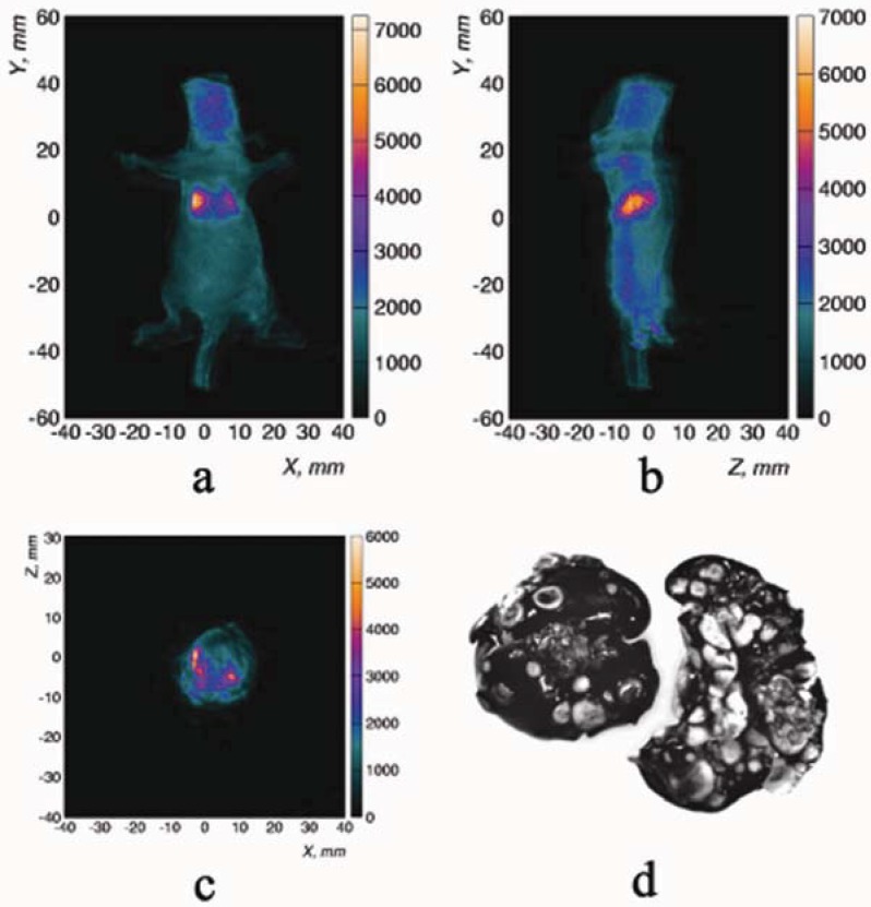
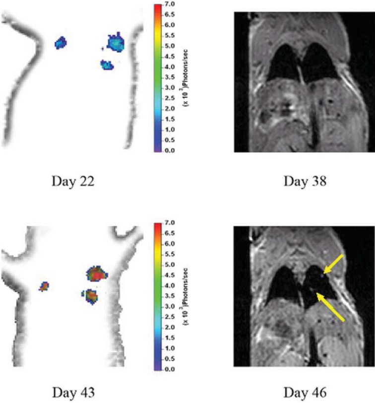
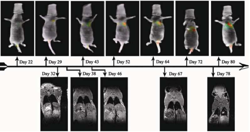
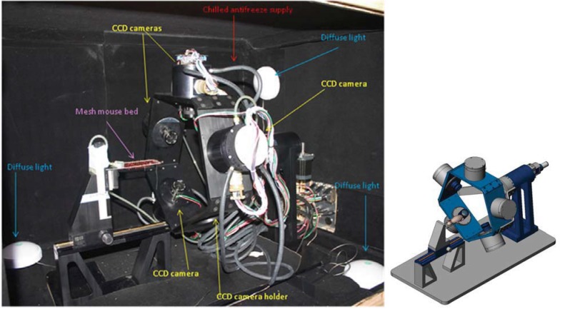
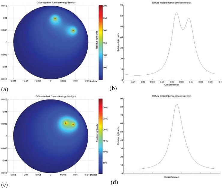
Bioluminescent imaging (BLI) of cells expressing luciferase is a valuable noninvasive technique for investigating molecular events and tumor dynamics in the living animal. Current usage is often limited to planar imaging, but tomographic imaging can enhance the usefulness of this technique in quantitative biomedical studies by allowing accurate determination of tumor size and attribution of the emitted light to a specific organ or tissue. Bioluminescence tomography based on a single camera with source rotation or mirrors to provide additional views has previously been reported. We report here in vivo studies using a novel approach with multiple rotating cameras that, when combined with image reconstruction software, provides the desired representation of point source metastases and other small lesions. Comparison with MRI validated the ability to detect lung tumor colonization in mouse lung.
生物发光成像是一种非常有价值的非侵入性技术,可用于活体动物体内分子事件和肿瘤动力学的研究。目前的应用通常仅限于平面成像,但断层成像可以通过准确确定肿瘤的大小并将发射的光归因于特定的器官或组织,从而增强该技术在定量生物医学研究中的有用性。基于带有旋转光源或镜子以提供附加视角的单个相机的生物发光断层成像先前已有报道。本文报道了一种新的使用多个旋转相机的方法的活体研究,该方法与图像重建软件相结合,可以对点源转移瘤和其他小病变进行所需的表示。与 MRI 的比较验证了检测小鼠肺部肿瘤定植的能力。