Liu Li, O'Kelly Devin, Schuetze Regan, Carlson Graham, Zhou Heling, Trawick Mary Lynn, Pinney Kevin G, Mason Ralph P
Department of Radiology, University of Texas Southwestern Medical Center, Dallas, TX 75390, USA.
Department of Chemistry and Biochemistry, Baylor University, Waco, TX 76798, USA.
Molecules. 2021 Apr 27;26(9):2551. doi: 10.3390/molecules26092551.
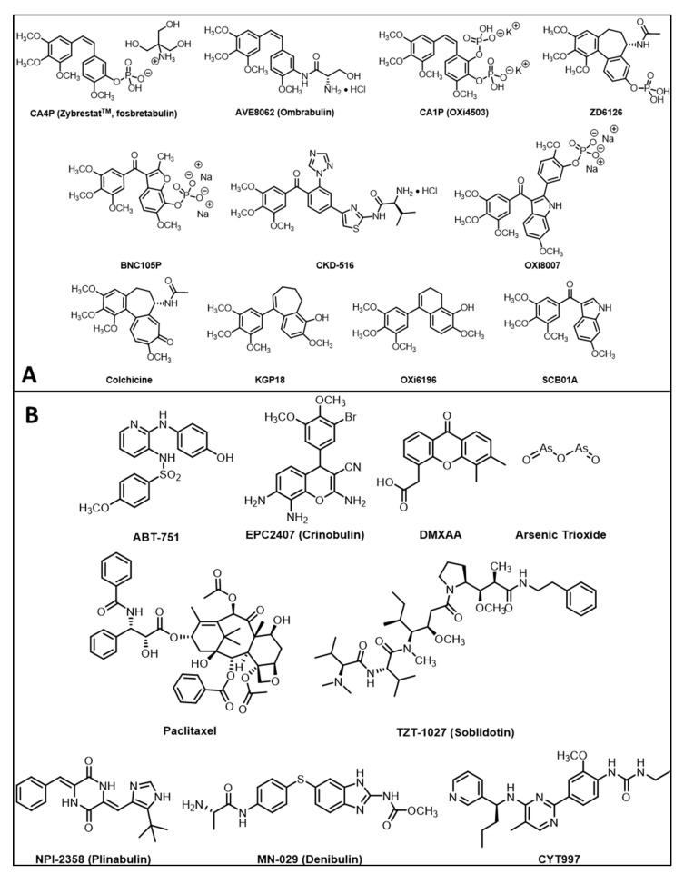
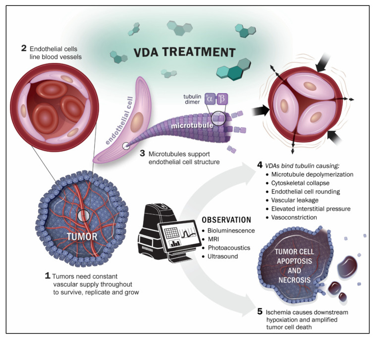
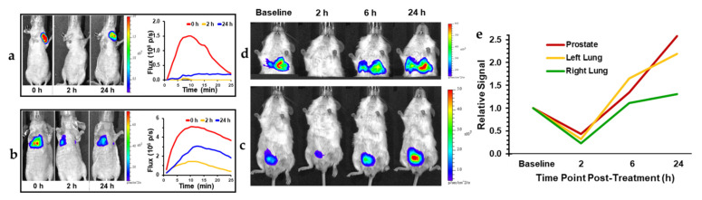
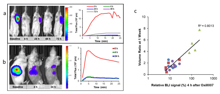
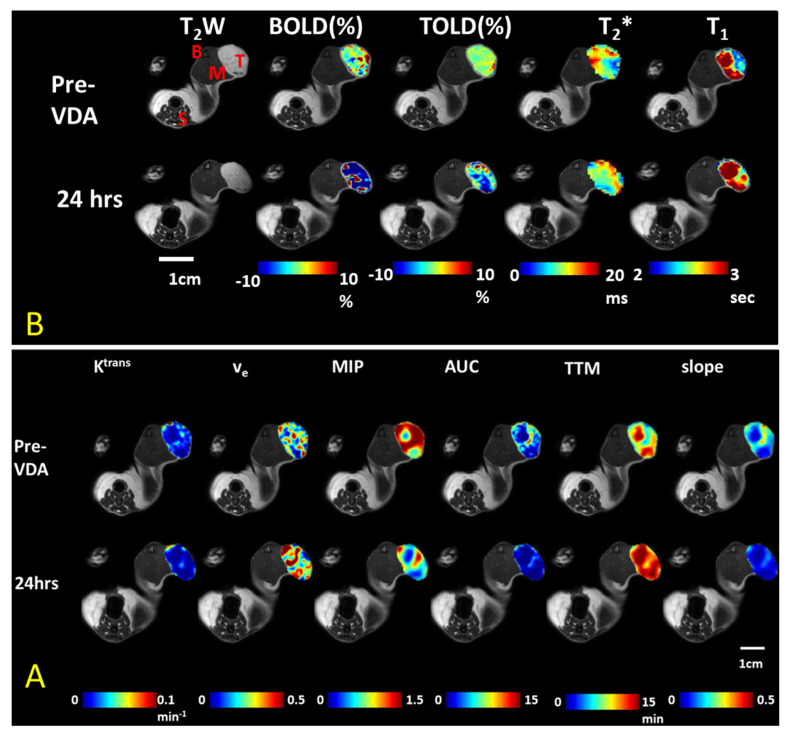
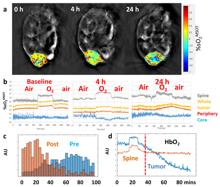
Tumor vasculature proliferates rapidly, generally lacks pericyte coverage, and is uniquely fragile making it an attractive therapeutic target. A subset of small-molecule tubulin binding agents cause disaggregation of the endothelial cytoskeleton leading to enhanced vascular permeability generating increased interstitial pressure. The resulting vascular collapse and ischemia cause downstream hypoxia, ultimately leading to cell death and necrosis. Thus, local damage generates massive amplification and tumor destruction. The tumor vasculature is readily accessed and potentially a common target irrespective of disease site in the body. Development of a therapeutic approach and particularly next generation agents benefits from effective non-invasive assays. Imaging technologies offer varying degrees of sophistication and ease of implementation. This review considers technological strengths and weaknesses with examples from our own laboratory. Methods reveal vascular extent and patency, as well as insights into tissue viability, proliferation and necrosis. Spatiotemporal resolution ranges from cellular microscopy to single slice tomography and full three-dimensional views of whole tumors and measurements can be sufficiently rapid to reveal acute changes or long-term outcomes. Since imaging is non-invasive, each tumor may serve as its own control making investigations particularly efficient and rigorous. The concept of tumor vascular disruption was proposed over 30 years ago and it remains an active area of research.
肿瘤血管迅速增殖,通常缺乏周细胞覆盖,且异常脆弱,这使其成为一个有吸引力的治疗靶点。一部分小分子微管蛋白结合剂会导致内皮细胞骨架解聚,从而增强血管通透性,使组织间隙压力升高。由此导致的血管塌陷和缺血会引起下游缺氧,最终导致细胞死亡和坏死。因此,局部损伤会引发大规模放大效应并导致肿瘤破坏。肿瘤血管易于触及,并且可能是一个通用靶点,而与身体中的疾病部位无关。开发一种治疗方法,尤其是新一代药物,受益于有效的非侵入性检测。成像技术的复杂程度和实施难度各不相同。本综述结合我们自己实验室的实例,探讨了这些技术的优缺点。这些方法可以揭示血管范围和通畅情况,以及对组织活力、增殖和坏死的洞察。时空分辨率范围从细胞显微镜检查到单层断层扫描,再到整个肿瘤的完整三维视图,测量速度足够快,可以揭示急性变化或长期结果。由于成像是非侵入性的,每个肿瘤都可以作为自身的对照,这使得研究特别高效和严谨。肿瘤血管破坏的概念在30多年前就已提出,它仍然是一个活跃的研究领域。