• 文献检索
  • 文档翻译
  • 深度研究
  • 学术资讯
  • Suppr Zotero 插件Zotero 插件
  • 邀请有礼
  • 套餐&价格
  • 历史记录
应用&插件
Suppr Zotero 插件Zotero 插件浏览器插件Mac 客户端Windows 客户端微信小程序
定价
高级版会员购买积分包购买API积分包
服务
文献检索文档翻译深度研究API 文档MCP 服务
关于我们
关于 Suppr公司介绍联系我们用户协议隐私条款
关注我们

Suppr 超能文献

核心技术专利:CN118964589B侵权必究
粤ICP备2023148730 号-1Suppr @ 2026

文献检索

告别复杂PubMed语法,用中文像聊天一样搜索,搜遍4000万医学文献。AI智能推荐,让科研检索更轻松。

立即免费搜索

文件翻译

保留排版,准确专业,支持PDF/Word/PPT等文件格式,支持 12+语言互译。

免费翻译文档

深度研究

AI帮你快速写综述,25分钟生成高质量综述,智能提取关键信息,辅助科研写作。

立即免费体验

在小鼠中,表达促黄体生成素释放激素mRNA的细胞源自嗅基板中的祖细胞的证据。

Evidence that cells expressing luteinizing hormone-releasing hormone mRNA in the mouse are derived from progenitor cells in the olfactory placode.

作者信息

Wray S, Grant P, Gainer H

机构信息

Laboratory of Neurochemistry, National Institute of Neurological Disorders and Stroke, Bethesda, MD 20892.

出版信息

Proc Natl Acad Sci U S A. 1989 Oct;86(20):8132-6. doi: 10.1073/pnas.86.20.8132.

DOI:10.1073/pnas.86.20.8132
PMID:2682637
原文链接:https://pmc.ncbi.nlm.nih.gov/articles/PMC298229/
Abstract

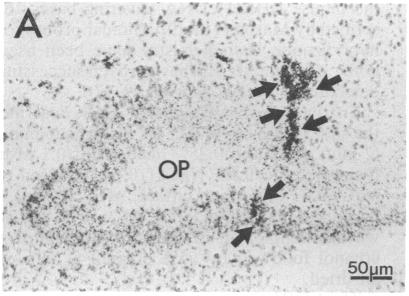
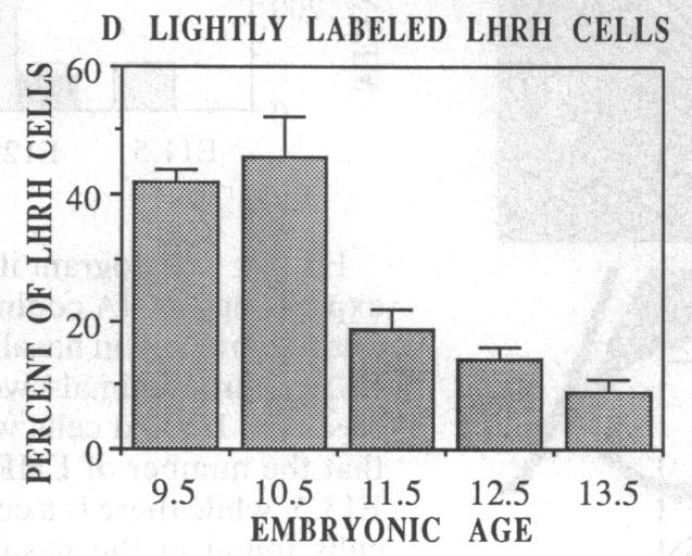
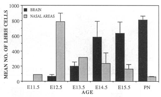
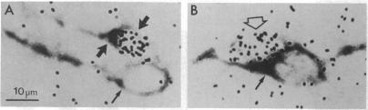
In situ hybridization histochemistry and immunocytochemistry were used to study the prenatal expression of luteinizing hormone-releasing hormone (LHRH) cells in the mouse. Cells expressing LHRH mRNA and peptide product were first detected on embryonic day 11.5 (E11.5) in the olfactory pit. On E12.5, the majority of LHRH cells were located on "tracks" extending from the olfactory pit to the base of the telencephalon. From E12.5 to E15.5, LHRH cells were detected in a rostral-to-caudal gradient in forebrain areas. Prior to E12.5, cells expressing LHRH mRNA were not detected in forebrain areas known to contain LHRH cells in postnatal animals. Quantitation of cells expressing LHRH mRNA showed that the number of labeled cells on E12.5 (approximately 800) equaled the number of LHRH cells in postnatal animals, but more than 90% of these cells were located in nasal regions. Between E12.5 and E15.5, the location of LHRH cells shifted. The number of LHRH cells in the forebrain increased, while the number of LHRH cells in nasal regions decreased over this same period. These findings establish that cells first found in the olfactory pit and thereafter in forebrain areas express the LHRH gene and correspond to the position of LHRH immunopositive cells found at these developmental times. To further examine the ontogeny of the LHRH system, immunocytochemistry in combination with [3H]thymidine autoradiography was used to determine when LHRH cells left the mitotic cycle. We show that LHRH neurons exhibit a discrete time of birth, suggesting that they arise as a single neuronal population between E10.0 and E11.0. Postnatal LHRH neurons were "birth-dated" shortly after differentiation of the olfactory placode and before LHRH mRNA was expressed in cells in the olfactory pit. Taken together, these studies support the hypothesis that all LHRH cells in the central nervous system arise from a discrete group of progenitor cells in the olfactory placode and that a subpopulation of these cells migrate into forebrain areas where they subsequently establish an adult-like distribution.

摘要

采用原位杂交组织化学和免疫细胞化学方法研究小鼠促黄体激素释放激素(LHRH)细胞的产前表达。在胚胎第11.5天(E11.5),首次在嗅窝中检测到表达LHRH mRNA和肽产物的细胞。在E12.5时,大多数LHRH细胞位于从嗅窝延伸至端脑底部的“轨迹”上。从E12.5到E15.5,在前脑区域按从前到后的梯度检测到LHRH细胞。在E12.5之前,在已知出生后动物中含有LHRH细胞的前脑区域未检测到表达LHRH mRNA的细胞。对表达LHRH mRNA的细胞进行定量分析表明,E12.5时标记细胞的数量(约800个)与出生后动物中LHRH细胞的数量相等,但这些细胞中90%以上位于鼻腔区域。在E12.5和E15.5之间,LHRH细胞的位置发生了变化。在此期间,前脑中LHRH细胞的数量增加,而鼻腔区域LHRH细胞的数量减少。这些发现证实,最初在嗅窝中发现、随后在前脑区域发现的细胞表达LHRH基因,并且与在这些发育阶段发现的LHRH免疫阳性细胞的位置相对应。为了进一步研究LHRH系统的个体发生,将免疫细胞化学与[3H]胸腺嘧啶放射自显影相结合,以确定LHRH细胞何时脱离有丝分裂周期。我们发现LHRH神经元具有离散的出生时间,这表明它们在E10.0至E11.0之间作为单个神经元群体出现。出生后的LHRH神经元在嗅基板分化后不久、且在嗅窝细胞中表达LHRH mRNA之前就已“确定出生时间”。综上所述,这些研究支持以下假说:中枢神经系统中的所有LHRH细胞均起源于嗅基板中一组离散的祖细胞,并且这些细胞的一个亚群迁移到前脑区域,随后在那里建立起类似成年动物的分布。

https://cdn.ncbi.nlm.nih.gov/pmc/blobs/fe0b/298229/6991f8dc825d/pnas00287-0484-b.jpg
https://cdn.ncbi.nlm.nih.gov/pmc/blobs/fe0b/298229/6c35ec2e8bdc/pnas00287-0482-a.jpg
https://cdn.ncbi.nlm.nih.gov/pmc/blobs/fe0b/298229/62095076be21/pnas00287-0482-b.jpg
https://cdn.ncbi.nlm.nih.gov/pmc/blobs/fe0b/298229/56e6beeef859/pnas00287-0482-c.jpg
https://cdn.ncbi.nlm.nih.gov/pmc/blobs/fe0b/298229/8fee07dcc4e1/pnas00287-0482-d.jpg
https://cdn.ncbi.nlm.nih.gov/pmc/blobs/fe0b/298229/1276fbca94a3/pnas00287-0482-e.jpg
https://cdn.ncbi.nlm.nih.gov/pmc/blobs/fe0b/298229/31217a89a5ff/pnas00287-0483-a.jpg
https://cdn.ncbi.nlm.nih.gov/pmc/blobs/fe0b/298229/75856543dacf/pnas00287-0483-b.jpg
https://cdn.ncbi.nlm.nih.gov/pmc/blobs/fe0b/298229/0c47cca44f4a/pnas00287-0484-a.jpg
https://cdn.ncbi.nlm.nih.gov/pmc/blobs/fe0b/298229/6991f8dc825d/pnas00287-0484-b.jpg

相似文献

1
Evidence that cells expressing luteinizing hormone-releasing hormone mRNA in the mouse are derived from progenitor cells in the olfactory placode.在小鼠中,表达促黄体生成素释放激素mRNA的细胞源自嗅基板中的祖细胞的证据。
Proc Natl Acad Sci U S A. 1989 Oct;86(20):8132-6. doi: 10.1073/pnas.86.20.8132.
2
Spatiotemporal cell expression of luteinizing hormone-releasing hormone in the prenatal mouse: evidence for an embryonic origin in the olfactory placode.促黄体生成素释放激素在产前小鼠中的时空细胞表达:嗅基板胚胎起源的证据。
Brain Res Dev Brain Res. 1989 Apr 1;46(2):309-18. doi: 10.1016/0165-3806(89)90295-2.
3
LHRH cells migrate on peripherin fibers in embryonic olfactory explant cultures: an in vitro model for neurophilic neuronal migration.促性腺激素释放激素(LHRH)细胞在胚胎嗅觉外植体培养物中的外周蛋白纤维上迁移:一种嗜神经性神经元迁移的体外模型。
Dev Biol. 1994 Nov;166(1):331-48. doi: 10.1006/dbio.1994.1319.
4
Two olfactory placode derived galanin subpopulations: luteinizing hormone-releasing hormone neurones and vomeronasal cells.源自嗅基板的两种甘丙肽亚群:促黄体生成素释放激素神经元和犁鼻器细胞。
J Neuroendocrinol. 2000 Jun;12(6):535-45. doi: 10.1046/j.1365-2826.2000.00486.x.
5
Ectopic expression of luteinizing hormone-releasing hormone and peripherin in the respiratory epithelium of mice lacking transcription factor AP-2alpha.缺乏转录因子AP-2α的小鼠呼吸上皮中促黄体生成激素释放激素和外周蛋白的异位表达。
Mech Dev. 2000 Jun;94(1-2):79-94. doi: 10.1016/s0925-4773(00)00316-6.
6
Deleted in colorectal cancer (DCC) regulates the migration of luteinizing hormone-releasing hormone neurons to the basal forebrain.结直肠癌缺失基因(DCC)调控促黄体生成素释放激素神经元向基底前脑的迁移。
J Neurosci. 2001 Feb 1;21(3):911-9. doi: 10.1523/JNEUROSCI.21-03-00911.2001.
7
Neurons that migrate from the olfactory epithelium in the chick express luteinizing hormone-releasing hormone.从鸡的嗅上皮迁移而来的神经元表达促黄体生成素释放激素。
Endocrinology. 1991 Mar;128(3):1676-8. doi: 10.1210/endo-128-3-1676.
8
Two populations of luteinizing hormone-releasing hormone neurons in the forebrain of the rhesus macaque during embryonic development.胚胎发育期间恒河猴前脑促黄体激素释放激素神经元的两种群体。
J Comp Neurol. 1997 Apr 14;380(3):293-309.
9
Direct evidence for the migration of LHRH neurons from the nasal region to the forebrain in the chick embryo: a carbocyanine dye analysis.鸡胚中促黄体激素释放激素(LHRH)神经元从鼻区迁移至前脑的直接证据:一种羰花青染料分析
Neurosci Res. 1994 May;19(3):331-8. doi: 10.1016/0168-0102(94)90046-9.
10
Midline nasal tissue influences nestin expression in nasal-placode-derived luteinizing hormone-releasing hormone neurons during development.在发育过程中,中线鼻组织影响源自鼻基板的促黄体生成激素释放激素神经元中的巢蛋白表达。
Dev Biol. 2000 Nov 15;227(2):343-57. doi: 10.1006/dbio.2000.9896.

引用本文的文献

1
Shared Lineage, Distinct Outcomes: Yap and Taz Loss Differentially Impact Schwann and Olfactory Ensheathing Cell Development Without Disrupting GnRH-1 Migration.共同谱系,不同结果:Yap和Taz缺失对施万细胞和嗅鞘细胞发育的影响不同,且不影响GnRH-1迁移。
Glia. 2025 Jul 9. doi: 10.1002/glia.70057.
2
Fibroblast growth factor 8: Multifaceted role in development and developmental disorder.成纤维细胞生长因子8:在发育及发育障碍中的多方面作用
Genes Dis. 2025 Jan 10;12(5):101524. doi: 10.1016/j.gendis.2025.101524. eCollection 2025 Sep.
3
Olfactory GnRH3 crypt sensory neurons transduce sex pheromone signals to induce male courtship behavior in zebrafish.

本文引用的文献

1
Time of vasopressin neuron origin in the mouse hypothalamus: examination by combined technique of immunocytochemistry and [3H]thymidine autoradiography.小鼠下丘脑加压素神经元的起源时间:免疫细胞化学与[3H]胸腺嘧啶核苷放射自显影联合技术检测
Brain Res. 1983 Aug;285(2):223-6. doi: 10.1016/0165-3806(83)90055-x.
2
[Disinfection of respiratory apparatus with formaldehyde in the aseptor (author's transl)].在无菌器中用甲醛对呼吸器械进行消毒(作者译)
Offentl Gesundheitswes. 1980 Feb;42(2):79-83.
3
Time of neuron origin in mouse hypothalamic nuclei.
嗅觉促性腺激素释放激素3(GnRH3)隐窝感觉神经元传导性信息素信号,以诱导斑马鱼的雄性求偶行为。
Sci China Life Sci. 2025 May 8. doi: 10.1007/s11427-025-2917-5.
4
Two cases of Kallmann syndrome caused by a novel mutation in ANOS1: A case report.两例由ANOS1基因新突变引起的卡尔曼综合征:病例报告。
Medicine (Baltimore). 2025 Apr 18;104(16):e42139. doi: 10.1097/MD.0000000000042139.
5
Intranasal kisspeptin administration rapidly stimulates gonadotropin release in humans.经鼻给予 kisspeptin 可迅速刺激人体促性腺激素释放。
EBioMedicine. 2025 May;115:105689. doi: 10.1016/j.ebiom.2025.105689. Epub 2025 Apr 11.
6
Transcriptomic profiling of murine GnRH neurons reveals developmental trajectories linked to human reproduction and infertility.小鼠促性腺激素释放激素神经元的转录组分析揭示了与人类生殖和不孕相关的发育轨迹。
Theranostics. 2025 Feb 26;15(8):3673-3692. doi: 10.7150/thno.91873. eCollection 2025.
7
Deletion in RMST lncRNA impairs hypothalamic neuronal development in a human stem cell-based model of Kallmann Syndrome.RMST长链非编码RNA的缺失在基于人干细胞的卡尔曼综合征模型中损害下丘脑神经元发育。
Cell Death Discov. 2024 Jul 19;10(1):330. doi: 10.1038/s41420-024-02074-4.
8
Anti-Müllerian Hormone: A Molecular Key to Unlocking Polycystic Ovary Syndrome?抗苗勒管激素:开启多囊卵巢综合征的分子钥匙?
Semin Reprod Med. 2024 Mar;42(1):41-48. doi: 10.1055/s-0044-1787525. Epub 2024 Jun 22.
9
Illuminating the terminal nerve: Uncovering the link between GnRH-1 neuron and olfactory development.揭示终神经:揭示 GnRH-1 神经元与嗅觉发育之间的联系。
J Comp Neurol. 2024 Mar;532(3):e25599. doi: 10.1002/cne.25599.
10
Cholinergic Control of GnRH Neuron Physiology and Luteinizing Hormone Secretion in Male Mice: Involvement of ACh/GABA Cotransmission.胆碱能控制 GnRH 神经元生理学和雄性小鼠促黄体激素分泌:涉及 ACh/GABA 共传递。
J Neurosci. 2024 Mar 20;44(12):e1780232024. doi: 10.1523/JNEUROSCI.1780-23.2024.
小鼠下丘脑核中神经元的起源时间。
Exp Neurol. 1973 Oct;41(1):163-73. doi: 10.1016/0014-4886(73)90187-8.
4
Neurophysin in the hypothalamo-neurohypophysial system. II. Immunocytochemical studies of the ontogeny of oxytocinergic and vasopressinergic neurons.下丘脑-神经垂体系统中的神经垂体素。II. 催产素能和加压素能神经元个体发生的免疫细胞化学研究。
J Neurosci. 1985 Jan;5(1):98-109. doi: 10.1523/JNEUROSCI.05-01-00098.1985.
5
Distribution of luteinizing hormone-releasing hormone in the nervus terminalis and brain of the mouse detected by immunocytochemistry.通过免疫细胞化学检测小鼠终神经和脑中促黄体生成激素释放激素的分布。
J Comp Neurol. 1987 Jan 8;255(2):231-44. doi: 10.1002/cne.902550207.
6
LHRH neurons in the female C57BL/6J mouse brain during reproductive aging: no loss up to middle age.雌性C57BL/6J小鼠大脑中促性腺激素释放激素(LHRH)神经元在生殖衰老过程中的变化:中年之前无损失。
Neurobiol Aging. 1986 Jan-Feb;7(1):45-8. doi: 10.1016/0197-4580(86)90026-6.
7
Development of the nervus terminalis in mammals including toothed whales and humans.包括齿鲸和人类在内的哺乳动物终神经的发育。
Ann N Y Acad Sci. 1987;519:447-64. doi: 10.1111/j.1749-6632.1987.tb36316.x.
8
Immunochemical studies of the location of luteinizing hormone-releasing hormone neurons in the nervus terminalis of the mouse.小鼠终神经中促黄体生成激素释放激素神经元定位的免疫化学研究。
Ann N Y Acad Sci. 1987;519:465-8. doi: 10.1111/j.1749-6632.1987.tb36317.x.
9
Specification of cerebral cortical areas.大脑皮质区域的特化
Science. 1988 Jul 8;241(4862):170-6. doi: 10.1126/science.3291116.
10
In situ hybridization histochemistry for messenger ribonucleic acid (mRNA) encoding gonadotropin-releasing hormone (GnRH): effect of estrogen on cellular levels of GnRH mRNA in female rat brain.编码促性腺激素释放激素(GnRH)的信使核糖核酸(mRNA)的原位杂交组织化学:雌激素对雌性大鼠脑内GnRH mRNA细胞水平的影响
Endocrinology. 1988 Jun;122(6):2570-7. doi: 10.1210/endo-122-6-2570.