Fisicaro Ryan A, Jost Ethan, Shaw Katharina, Brennan Nicole Petrovich, Peck Kyung K, Holodny Andrei I
*Functional MRI Laboratory †Department of Radiology ‡Brain Tumor Canter §Department of Medical Physics, Memorial Sloan-Kettering Cancer Center, New York ¶Department of Psychology, Cornell University, Ithaca, NY.
Top Magn Reson Imaging. 2016 Feb;25(1):25-30. doi: 10.1097/RMR.0000000000000077.
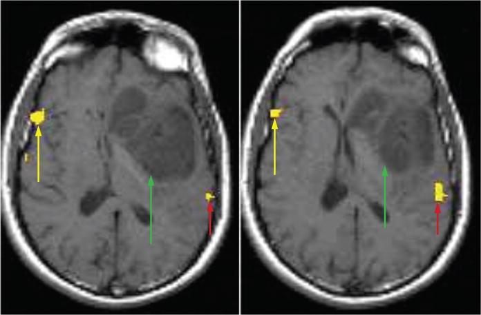
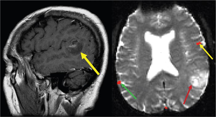
Cortical reorganization of function due to the growth of an adjacent brain tumor has clearly been demonstrated in a number of surgically proven cases. Such cases demonstrate the unmistakable implications for the neurosurgical treatment of brain tumors, as the cortical function may not reside where one may initially suspect based solely on the anatomical magnetic resonance imaging (MRI). Consequently, preoperative localization of eloquent areas adjacent to a brain tumor is necessary, as this may demonstrate unexpected organization, which may affect the neurosurgical approach to the lesion. However, in interpreting functional MRI studies, the interpreting physician must be cognizant of artifacts, which may limit the accuracy of functional MRI in the setting of brain tumors.
在一些经手术证实的病例中,已明确显示出由于相邻脑肿瘤生长导致的皮质功能重组。此类病例表明了对脑肿瘤神经外科治疗有着明确无误的影响,因为皮质功能可能并不在仅基于解剖磁共振成像(MRI)最初所怀疑的位置。因此,术前对脑肿瘤相邻的明确功能区进行定位是必要的,因为这可能显示出意想不到的组织情况,而这可能会影响对病变的神经外科手术方法。然而,在解读功能磁共振成像研究时,解读医生必须认识到伪影,在脑肿瘤情况下,伪影可能会限制功能磁共振成像的准确性。