Sillerud Laurel O
BRaIN Center, Department of Neurology, University of New Mexico School of Medicine, Albuquerque, NM, USA.
Int J Nanomedicine. 2016 Jan 20;11:357-71. doi: 10.2147/IJN.S93409. eCollection 2016.
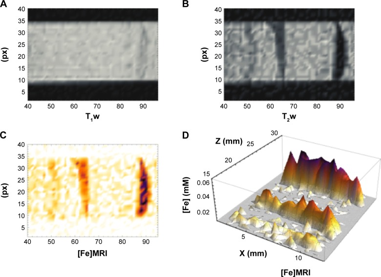
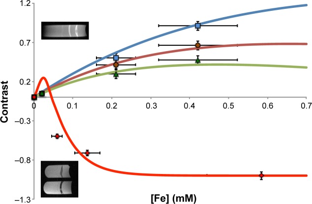
We report the development, experimental verification, and application of a general theory called [Fe]MRI (pronounced fem-ree) for the non-invasive, quantitative molecular magnetic resonance imaging (MRI) of added magnetic nanoparticles or other magnetic contrast agents in biological tissues and other sites. [Fe]MRI can easily be implemented on any MRI instrument, requiring only measurements of the background nuclear magnetic relaxation times (T1, T2) of the tissue of interest, injection of the magnetic particles, and the subsequent acquisition of a pair of T1-weighted and T2-weighted images. These images, converted into contrast images, are subtracted to yield a contrast difference image proportional to the absolute nanoparticle, iron concentration, ([Fe]) image. [Fe]MRI was validated with the samples of superparamagnetic iron oxide nanoparticles (SPIONs) both in agarose gels and bound to human prostate tumor cells. The [Fe]MRI measurement of the binding of anti-prostate specific membrane antigen (PSMA) conjugated SPIONs to PSMA-positive LNCaP and PSMA-negative DU145 cells in vitro allowed a facile discrimination among prostate tumor cell types based on their PSMA expression level. The low [Fe] detection limit of ~2 μM for SPIONs allows sensitive MRI of added iron at concentrations considerably below the US Food and Drug Administration's human iron dosage guidelines (<90 μM, 5 mg/kg).
我们报告了一种名为[Fe]MRI(发音为fem-ree)的通用理论的开发、实验验证及应用,该理论用于对生物组织及其他部位中添加的磁性纳米颗粒或其他磁性造影剂进行非侵入性定量分子磁共振成像(MRI)。[Fe]MRI可轻松在任何MRI仪器上实现,只需测量感兴趣组织的背景核磁弛豫时间(T1、T2),注射磁性颗粒,随后采集一对T1加权和T2加权图像。将这些图像转换为对比图像后相减,以生成与绝对纳米颗粒铁浓度([Fe])图像成比例的对比差异图像。[Fe]MRI在琼脂糖凝胶中和与人前列腺肿瘤细胞结合的超顺磁性氧化铁纳米颗粒(SPIONs)样本上得到了验证。[Fe]MRI对体外抗前列腺特异性膜抗原(PSMA)偶联的SPIONs与PSMA阳性LNCaP和PSMA阴性DU145细胞结合的测量,使得基于前列腺肿瘤细胞类型的PSMA表达水平能够轻松区分。SPIONs约2μM的低[Fe]检测限允许对添加铁进行敏感的MRI检测,其浓度远低于美国食品药品监督管理局的人体铁剂量指南(<90μM,5mg/kg)。