Wei Shu, Tao Wei, Zhu Hongwei, Li Yongjie
Xuanwu Hospital, Capital Medical University, Beijing, China.
Wideochir Inne Tech Maloinwazyjne. 2016 Jan;10(4):555-60. doi: 10.5114/wiitm.2015.55845. Epub 2015 Nov 27.
Successful percutaneous endoscopic lumbar discectomy (PELD) requires an appropriate working trajectory. Due to the complexity of spinal anatomy, this is difficult to verify with conventional 2-dimensional fluoroscopy.
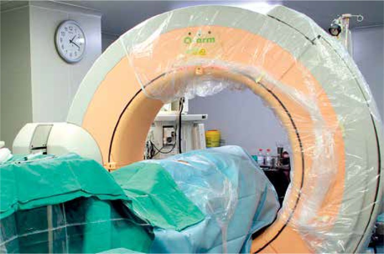
Here we assessed the feasibility and utility of the O-arm for establishing the working trajectory for PELD.
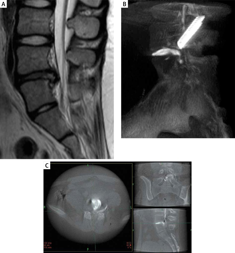
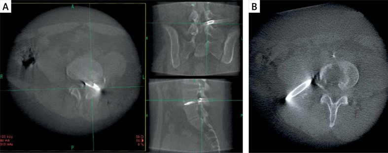
We retrospectively reviewed the records of 89 patients with lumbar disc herniation who underwent PELD using the O-arm. The working trajectory was evaluated with standard fluoroscopic images or 3-dimensional, volumetric computed tomography scan. Based on the detail provided by the multiplanar view, we confirmed the ideal working trajectory and adjusted the surgical approach accordingly.
At the 12-month follow-up, based on MacNab's criteria, 85.9% of patients showed an excellent or good outcome. There were no major complications.
The O-arm provides detailed multiplanar intraoperative high-quality imaging for PELD, and enables the surgeon to ascertain the surgical anatomy, determine the optimal working trajectory, and improve the accuracy of surgery.
成功的经皮内镜下腰椎间盘切除术(PELD)需要合适的工作通道。由于脊柱解剖结构的复杂性,使用传统的二维荧光透视很难进行验证。
在此,我们评估了O型臂用于建立PELD工作通道的可行性和实用性。
我们回顾性分析了89例行PELD并使用O型臂的腰椎间盘突出症患者的记录。通过标准荧光透视图像或三维容积计算机断层扫描来评估工作通道。根据多平面视图提供的细节,我们确定了理想的工作通道并相应地调整了手术入路。
在12个月的随访中,根据MacNab标准,85.9%的患者显示出优或良的结果。无重大并发症。
O型臂为PELD提供了详细的多平面术中高质量成像,使外科医生能够确定手术解剖结构,确定最佳工作通道,并提高手术准确性。