• 文献检索
  • 文档翻译
  • 深度研究
  • 学术资讯
  • Suppr Zotero 插件Zotero 插件
  • 邀请有礼
  • 套餐&价格
  • 历史记录
应用&插件
Suppr Zotero 插件Zotero 插件浏览器插件Mac 客户端Windows 客户端微信小程序
定价
高级版会员购买积分包购买API积分包
服务
文献检索文档翻译深度研究API 文档MCP 服务
关于我们
关于 Suppr公司介绍联系我们用户协议隐私条款
关注我们

Suppr 超能文献

核心技术专利:CN118964589B侵权必究
粤ICP备2023148730 号-1Suppr @ 2026

文献检索

告别复杂PubMed语法,用中文像聊天一样搜索,搜遍4000万医学文献。AI智能推荐,让科研检索更轻松。

立即免费搜索

文件翻译

保留排版,准确专业,支持PDF/Word/PPT等文件格式,支持 12+语言互译。

免费翻译文档

深度研究

AI帮你快速写综述,25分钟生成高质量综述,智能提取关键信息,辅助科研写作。

立即免费体验

冠状动脉内成像在易损斑块检测中的应用

Intracoronary Imaging in the Detection of Vulnerable Plaques.

作者信息

Batty Jonathan A, Subba Shristy, Luke Peter, Gigi Li Wing Chi, Sinclair Hannah, Kunadian Vijay

机构信息

Institute of Cellular Medicine, Newcastle University, 3rd Floor, William Leech Building, Newcastle Upon Tyne, NE2 4HH, UK.

Freeman Hospital, Newcastle Upon Tyne NHS Foundation Trust, Newcastle Upon Tyne, NE7 7DN, UK.

出版信息

Curr Cardiol Rep. 2016 Mar;18(3):28. doi: 10.1007/s11886-016-0705-1.

DOI:10.1007/s11886-016-0705-1
PMID:26879196
原文链接:https://pmc.ncbi.nlm.nih.gov/articles/PMC4754333/
Abstract

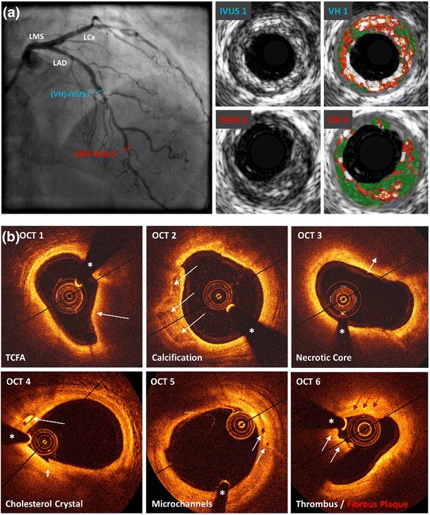
Coronary artery disease is the result of atherosclerotic changes to the coronary arterial wall, comprising endothelial dysfunction, vascular inflammation and deposition of lipid-rich macrophage foam cells. Certain high-risk atherosclerotic plaques are vulnerable to disruption, leading to rupture, thrombosis and the clinical sequelae of acute coronary syndrome. Though recognised as the gold standard for evaluating the presence, distribution and severity of atherosclerotic lesions, invasive coronary angiography is incapable of identifying non-stenotic, vulnerable plaques that are responsible for adverse cardiovascular events. The recognition of such limitations has impelled the development of intracoronary imaging technologies, including intravascular ultrasound, optical coherence tomography and near-infrared spectroscopy, which enable the detailed evaluation of the coronary wall and atherosclerotic plaques in clinical practice. This review discusses the present status of invasive imaging technologies; summarises up-to-date, evidence-based clinical guidelines; and addresses questions that remain unanswered with regard to the future of intracoronary plaque imaging.

摘要

冠状动脉疾病是冠状动脉壁发生动脉粥样硬化改变的结果,包括内皮功能障碍、血管炎症以及富含脂质的巨噬细胞泡沫细胞的沉积。某些高危动脉粥样硬化斑块易发生破裂,导致急性冠状动脉综合征的临床后遗症,如破裂、血栓形成等。尽管有创冠状动脉造影被公认为评估动脉粥样硬化病变的存在、分布和严重程度的金标准,但它无法识别导致不良心血管事件的非狭窄性易损斑块。对这些局限性的认识推动了冠状动脉内成像技术的发展,包括血管内超声、光学相干断层扫描和近红外光谱,这些技术能够在临床实践中对冠状动脉壁和动脉粥样硬化斑块进行详细评估。本综述讨论了有创成像技术的现状;总结了最新的、基于证据的临床指南;并探讨了冠状动脉斑块成像未来仍未得到解答的问题。

https://cdn.ncbi.nlm.nih.gov/pmc/blobs/c1a8/4754333/782c631c5fa7/11886_2016_705_Fig1_HTML.jpg

相似文献

1
Intracoronary Imaging in the Detection of Vulnerable Plaques.冠状动脉内成像在易损斑块检测中的应用
Curr Cardiol Rep. 2016 Mar;18(3):28. doi: 10.1007/s11886-016-0705-1.
2
Targeted Near-Infrared Fluorescence Imaging of Atherosclerosis: Clinical and Intracoronary Evaluation of Indocyanine Green.动脉粥样硬化的靶向近红外荧光成像:吲哚菁绿的临床及冠状动脉内评估
JACC Cardiovasc Imaging. 2016 Sep;9(9):1087-1095. doi: 10.1016/j.jcmg.2016.01.034. Epub 2016 Aug 17.
3
Near-infrared spectroscopy for intracoronary detection of lipid-rich plaques to understand atherosclerotic plaque biology in man and guide clinical therapy.近红外光谱技术用于冠状动脉内脂质丰富斑块的检测,以了解人类动脉粥样硬化斑块的生物学特性,并指导临床治疗。
J Intern Med. 2015 Aug;278(2):110-25. doi: 10.1111/joim.12381. Epub 2015 Jun 19.
4
Imaging of the vulnerable plaque: new modalities.易损斑块的影像学检查:新方法
South Med J. 2003 Nov;96(11):1142-7. doi: 10.1097/01.SMJ.0000089063.76530.04.
5
The Role of Intracoronary Plaque Imaging with Intravascular Ultrasound, Optical Coherence Tomography, and Near-Infrared Spectroscopy in Patients with Coronary Artery Disease.冠状动脉内斑块成像(血管内超声、光学相干断层扫描和近红外光谱)在冠状动脉疾病患者中的作用。
Curr Atheroscler Rep. 2016 Sep;18(9):57. doi: 10.1007/s11883-016-0607-0.
6
Invasive imaging of the coronary atherosclerotic plaque.冠状动脉粥样硬化斑块的侵入性成像。
Minerva Cardioangiol. 2012 Jun;60(3):305-29.
7
Intravascular modalities for detection of vulnerable plaque: current status.用于检测易损斑块的血管内技术:现状
Arterioscler Thromb Vasc Biol. 2003 Aug 1;23(8):1333-42. doi: 10.1161/01.ATV.0000080948.08888.BF. Epub 2003 Jun 12.
8
Invasive testing for coronary artery disease: FFR, IVUS, OCT, NIRS.冠状动脉疾病的侵入性检测:血流储备分数(FFR)、血管内超声(IVUS)、光学相干断层扫描(OCT)、近红外光谱(NIRS)。
Cardiol Clin. 2014 Aug;32(3):405-17. doi: 10.1016/j.ccl.2014.04.005.
9
[Invasive methods of detection of unstable atherosclerotic plaques in coronary arteries].[冠状动脉不稳定动脉粥样硬化斑块的侵入性检测方法]
Kardiologiia. 2014;54(11):46-56. doi: 10.18565/cardio.2014.11.46-56.
10
Identification of patients and plaques vulnerable to future coronary events with near-infrared spectroscopy intravascular ultrasound imaging: a prospective, cohort study.应用近红外光谱血管内超声成像技术识别易发生未来冠状动脉事件的患者和斑块:一项前瞻性队列研究。
Lancet. 2019 Nov 2;394(10209):1629-1637. doi: 10.1016/S0140-6736(19)31794-5. Epub 2019 Sep 27.

引用本文的文献

1
Reporting trends and clinical impact of high-risk coronary plaques on cardiac CT angiography in CAD-RADS.CAD-RADS中高危冠状动脉斑块在心脏CT血管造影上的报告趋势及临床影响
Eur Radiol. 2025 Jul 30. doi: 10.1007/s00330-025-11872-y.
2
Intracoronary Imaging of Vulnerable Plaque-From Clinical Research to Everyday Practice.易损斑块的冠状动脉内成像——从临床研究到日常实践
J Clin Med. 2022 Nov 9;11(22):6639. doi: 10.3390/jcm11226639.
3
Application of Non-invasive Imaging in Inflammatory Disease Conditions to Evaluate Subclinical Coronary Artery Disease.

本文引用的文献

1
Optical coherence tomography to evaluate coronary stent implantation and complications.光学相干断层扫描评估冠状动脉支架植入及并发症
Coron Artery Dis. 2015 Aug;26 Suppl 1:e55-68. doi: 10.1097/MCA.0000000000000215.
2
Role of near-infrared spectroscopy in intravascular coronary imaging.近红外光谱在血管内冠状动脉成像中的作用。
Cardiovasc Revasc Med. 2015 Jul-Aug;16(5):299-305. doi: 10.1016/j.carrev.2015.06.001. Epub 2015 Jun 10.
3
The basics of intravascular optical coherence tomography.血管内光学相干断层扫描基础
无创影像学在炎症性疾病条件下评估亚临床冠状动脉疾病中的应用。
Curr Rheumatol Rep. 2019 Dec 12;22(1):1. doi: 10.1007/s11926-019-0875-0.
4
Imaging Cardiovascular Calcification.心血管钙化成像
J Am Heart Assoc. 2018 Jun 28;7(13):e008564. doi: 10.1161/JAHA.118.008564.
5
Association of D-dimer with Plaque Characteristics and Plasma Biomarkers of Oxidation-Specific Epitopes in Stable Subjects with Coronary Artery Disease.D-二聚体与稳定型冠心病患者斑块特征及氧化特异性表位血浆生物标志物的相关性。
J Cardiovasc Transl Res. 2018 Jun;11(3):221-229. doi: 10.1007/s12265-018-9790-4. Epub 2018 Jan 17.
6
Coronary Plaque Microstructure and Composition Modify Optical Polarization: A New Endogenous Contrast Mechanism for Optical Frequency Domain Imaging.冠状动脉斑块的微观结构和组成改变光的偏振:光频域成像的一种新的内源性对比机制。
JACC Cardiovasc Imaging. 2018 Nov;11(11):1666-1676. doi: 10.1016/j.jcmg.2017.09.023. Epub 2017 Dec 13.
Postepy Kardiol Interwencyjnej. 2015;11(2):74-83. doi: 10.5114/pwki.2015.52278. Epub 2015 Jun 20.
4
Serial Optical Coherence Tomography Evaluation at 6, 12, and 24 Months After Biolimus A9-Eluting Biodegradable Polymer-Coated Stent Implantation.生物可吸收聚合物涂层雷帕霉素洗脱支架置入后 6、12 和 24 个月的连续光学相干断层扫描评估
Can J Cardiol. 2015 Aug;31(8):980-8. doi: 10.1016/j.cjca.2015.02.036. Epub 2015 Mar 5.
5
Multimodality Intravascular Imaging to Predict Periprocedural Myocardial Infarction During Percutaneous Coronary Intervention.多模态血管内影像学预测经皮冠状动脉介入治疗围术期心肌梗死。
JACC Cardiovasc Interv. 2015 Jun;8(7):937-45. doi: 10.1016/j.jcin.2015.03.016.
6
Plaque Characterization to Inform the Prediction and Prevention of Periprocedural Myocardial Infarction During Percutaneous Coronary Intervention: The CANARY Trial (Coronary Assessment by Near-infrared of Atherosclerotic Rupture-prone Yellow).基于近红外光检测易损斑块对经皮冠状动脉介入治疗围术期心肌梗死的预测与预防作用的研究:CANARY 试验(基于近红外光技术检测易损斑块的粥样硬化破裂倾向的黄色研究)。
JACC Cardiovasc Interv. 2015 Jun;8(7):927-36. doi: 10.1016/j.jcin.2015.01.032. Epub 2015 May 20.
7
Assessment of angiographic coronary calcification and plaque composition in virtual histology intravascular ultrasound.虚拟组织学血管内超声对冠状动脉造影钙化和斑块成分的评估。
J Interv Cardiol. 2015 Apr;28(2):205-14. doi: 10.1111/joic.12189. Epub 2015 Apr 2.
8
OCT for the identification of vulnerable plaque in acute coronary syndrome.OCT 用于急性冠脉综合征中易损斑块的识别。
JACC Cardiovasc Imaging. 2015 Feb;8(2):198-209. doi: 10.1016/j.jcmg.2014.12.005.
9
Combined IVUS and NIRS detection of fibroatheromas: histopathological validation in human coronary arteries.联合 IVUS 和 NIRS 检测纤维脂肪斑:人体冠状动脉的组织病理学验证。
JACC Cardiovasc Imaging. 2015 Feb;8(2):184-94. doi: 10.1016/j.jcmg.2014.09.021. Epub 2015 Jan 7.
10
Heart disease and stroke statistics--2015 update: a report from the American Heart Association.《2015年心脏病和中风统计数据更新:美国心脏协会报告》
Circulation. 2015 Jan 27;131(4):e29-322. doi: 10.1161/CIR.0000000000000152. Epub 2014 Dec 17.