Ong Ta-Hsuan, Romanova Elena V, Roberts-Galbraith Rachel H, Yang Ning, Zimmerman Tyler A, Collins James J, Lee Ji Eun, Kelleher Neil L, Newmark Phillip A, Sweedler Jonathan V
From the Department of Chemistry, and the Beckman Institute.
the Department of Cell and Developmental Biology, Howard Hughes Medical Institute, University of Illinois at Urbana-Champaign, Urbana, Illinois 61801, and.
J Biol Chem. 2016 Apr 8;291(15):8109-20. doi: 10.1074/jbc.M115.709196. Epub 2016 Feb 16.
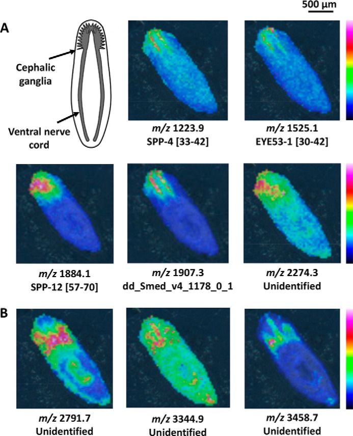
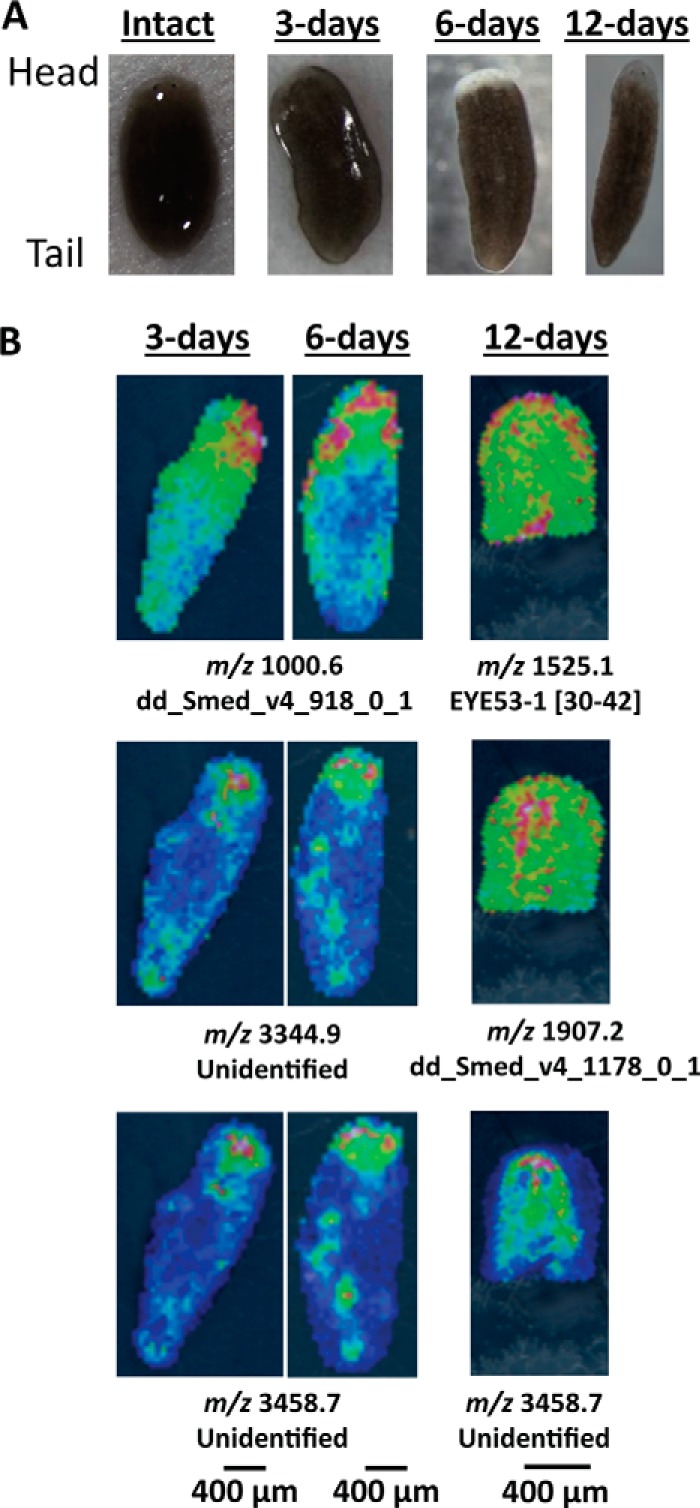
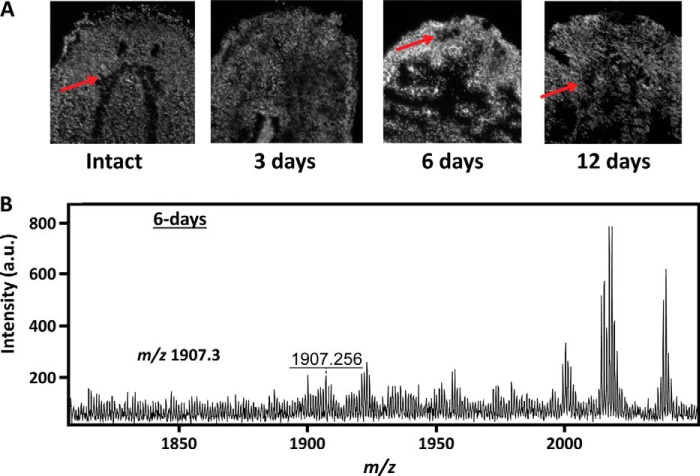
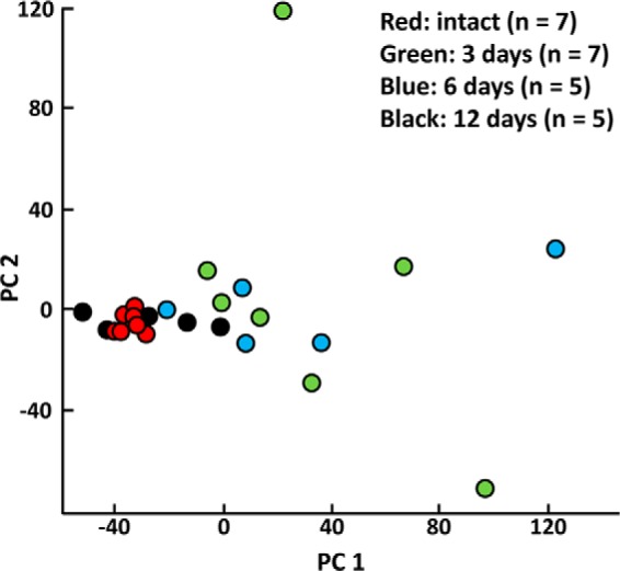
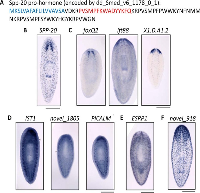
Tissue regeneration is a complex process that involves a mosaic of molecules that vary spatially and temporally. Insights into the chemical signaling underlying this process can be achieved with a multiplex and untargeted chemical imaging method such as mass spectrometry imaging (MSI), which can enablede novostudies of nervous system regeneration. A combination of MSI and multivariate statistics was used to differentiate peptide dynamics in the freshwater planarian flatwormSchmidtea mediterraneaat different time points during cephalic ganglia regeneration. A protocol was developed to makeS. mediterraneatissues amenable for MSI. MS ion images of planarian tissue sections allow changes in peptides and unknown compounds to be followed as a function of cephalic ganglia regeneration. In conjunction with fluorescence imaging, our results suggest that even though the cephalic ganglia structure is visible after 6 days of regeneration, the original chemical composition of these regenerated structures is regained only after 12 days. Differences were observed in many peptides, such as those derived from secreted peptide 4 and EYE53-1. Peptidomic analysis further identified multiple peptides from various known prohormones, histone proteins, and DNA- and RNA-binding proteins as being associated with the regeneration process. Mass spectrometry data also facilitated the identification of a new prohormone, which we have named secreted peptide prohormone 20 (SPP-20), and is up-regulated during regeneration in planarians.
组织再生是一个复杂的过程,涉及到在空间和时间上变化的多种分子。通过质谱成像(MSI)等多重且非靶向的化学成像方法,可以深入了解这一过程背后的化学信号传导,该方法能够实现对神经系统再生的全新研究。MSI与多变量统计相结合,用于区分淡水涡虫地中海涡虫(Schmidtea mediterranea)在头神经节再生过程中不同时间点的肽动态变化。开发了一种方案,使地中海涡虫组织适用于MSI。涡虫组织切片的MS离子图像能够跟踪肽和未知化合物随头神经节再生的变化。结合荧光成像,我们的结果表明,尽管再生6天后头神经节结构可见,但这些再生结构的原始化学成分仅在12天后才恢复。在许多肽中观察到差异,例如那些源自分泌肽4和EYE53-1的肽。肽组学分析进一步鉴定出多种来自各种已知前体激素、组蛋白以及DNA和RNA结合蛋白的肽与再生过程相关。质谱数据还有助于鉴定一种新前体激素,我们将其命名为分泌肽前体激素20(SPP-20),它在涡虫再生过程中上调。