Riccio Paul, Cebrian Cristina, Zong Hui, Hippenmeyer Simon, Costantini Frank
Department of Genetics and Development, Columbia University, New York, New York, United States of America.
Department of Microbiology, Immunology, and Cancer Biology, University of Virginia School of Medicine, Charlottesville, Virginia, United States of America.
PLoS Biol. 2016 Feb 19;14(2):e1002382. doi: 10.1371/journal.pbio.1002382. eCollection 2016 Feb.
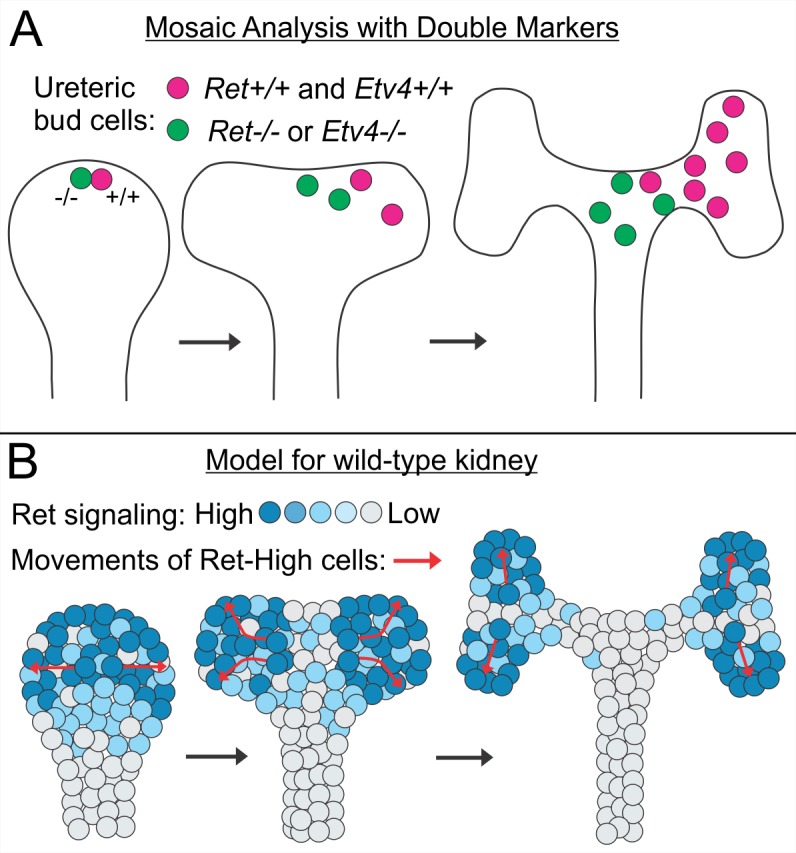
Branching morphogenesis of the epithelial ureteric bud forms the renal collecting duct system and is critical for normal nephron number, while low nephron number is implicated in hypertension and renal disease. Ureteric bud growth and branching requires GDNF signaling from the surrounding mesenchyme to cells at the ureteric bud tips, via the Ret receptor tyrosine kinase and coreceptor Gfrα1; Ret signaling up-regulates transcription factors Etv4 and Etv5, which are also critical for branching. Despite extensive knowledge of the genetic control of these events, it is not understood, at the cellular level, how renal branching morphogenesis is achieved or how Ret signaling influences epithelial cell behaviors to promote this process. Analysis of chimeric embryos previously suggested a role for Ret signaling in promoting cell rearrangements in the nephric duct, but this method was unsuited to study individual cell behaviors during ureteric bud branching. Here, we use Mosaic Analysis with Double Markers (MADM), combined with organ culture and time-lapse imaging, to trace the movements and divisions of individual ureteric bud tip cells. We first examine wild-type clones and then Ret or Etv4 mutant/wild-type clones in which the mutant and wild-type sister cells are differentially and heritably marked by green and red fluorescent proteins. We find that, in normal kidneys, most individual tip cells behave as self-renewing progenitors, some of whose progeny remain at the tips while others populate the growing UB trunks. In Ret or Etv4 MADM clones, the wild-type cells generated at a UB tip are much more likely to remain at, or move to, the new tips during branching and elongation, while their Ret-/- or Etv4-/- sister cells tend to lag behind and contribute only to the trunks. By tracking successive mitoses in a cell lineage, we find that Ret signaling has little effect on proliferation, in contrast to its effects on cell movement. Our results show that Ret/Etv4 signaling promotes directed cell movements in the ureteric bud tips, and suggest a model in which these cell movements mediate branching morphogenesis.
上皮输尿管芽的分支形态发生形成了肾集合管系统,对正常肾单位数量至关重要,而肾单位数量少与高血压和肾脏疾病有关。输尿管芽的生长和分支需要周围间充质通过Ret受体酪氨酸激酶和共受体Gfrα1向输尿管芽尖端的细胞传递GDNF信号;Ret信号上调转录因子Etv4和Etv5,它们对分支也很关键。尽管对这些事件的遗传控制有广泛了解,但在细胞水平上,尚不清楚肾分支形态发生是如何实现的,以及Ret信号如何影响上皮细胞行为以促进这一过程。对嵌合胚胎的分析先前表明Ret信号在促进肾管中的细胞重排中起作用,但这种方法不适合研究输尿管芽分支过程中的单个细胞行为。在这里,我们使用双标记镶嵌分析(MADM),结合器官培养和延时成像,来追踪单个输尿管芽尖端细胞的运动和分裂。我们首先检查野生型克隆,然后检查Ret或Etv4突变体/野生型克隆,其中突变体和野生型姐妹细胞通过绿色和红色荧光蛋白进行差异和可遗传标记。我们发现,在正常肾脏中,大多数单个尖端细胞表现为自我更新的祖细胞,其一些后代留在尖端,而另一些则填充生长中的输尿管芽主干。在Ret或Etv4 MADM克隆中,在输尿管芽尖端产生的野生型细胞在分支和伸长过程中更有可能留在或移动到新的尖端,而它们的Ret-/-或Etv4-/-姐妹细胞往往落后,仅对主干有贡献。通过追踪细胞谱系中的连续有丝分裂,我们发现Ret信号对增殖影响很小,这与其对细胞运动的影响形成对比。我们的结果表明,Ret/Etv4信号促进输尿管芽尖端的定向细胞运动,并提出了一个模型,其中这些细胞运动介导分支形态发生。