Oglesby Ericka N, Tezel Gülgün, Cone-Kimball Elizabeth, Steinhart Matthew R, Jefferys Joan, Pease Mary E, Quigley Harry A
Glaucoma Center of Excellence, Wilmer Ophthalmological Institute Department of Ophthalmology, Johns Hopkins University, Baltimore, MD.
Department of Ophthalmology, Columbia University, New York, NY.
Mol Vis. 2016 Jan 29;22:82-99. eCollection 2016.
To study the detailed cellular and molecular changes in the mouse sclera subjected to experimental glaucoma.
Three strains of mice underwent experimental bead-injection glaucoma and were euthanized at 3 days and 1, 3, and 6 weeks. Scleral protein expression was analyzed with liquid chromatography coupled with tandem mass spectrometry (LC-MS/MS) using (16)O/(18)O labeling for quantification in 1- and 6-week tissues. Sclera protein samples were also analyzed with immunoblotting with specific antibodies to selected proteins. The proportion of proliferating scleral fibroblasts was quantified with Ki67 and 4',6-diamidino-2-phenylindole (DAPI) labeling, and selected proteins were studied with immunohistochemistry.
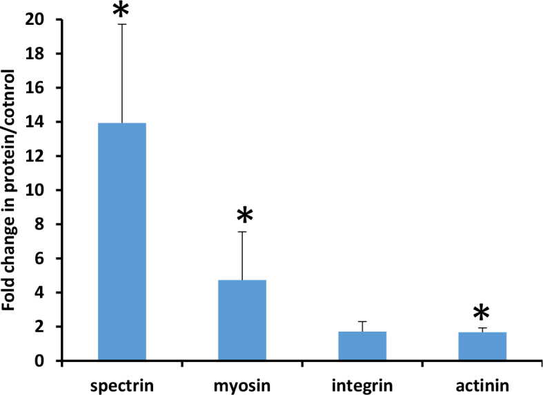
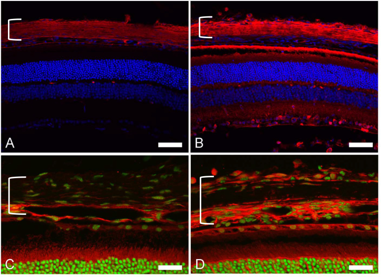
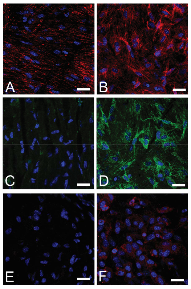
Proteomic analysis showed increases in molecules involved in integrin-linked kinase signaling and actin cytoskeleton signaling pathways at 1 and 6 weeks after experimental glaucoma. The peripapillary scleral region had more fibroblasts than equatorial sclera (p=0.001, n=217, multivariable regression models). There was a sixfold increase in proliferating fibroblasts in the experimental glaucoma sclera at 1 week and a threefold rise at 3 and 6 weeks (p=0.0005, univariate regression). Immunoblots confirmed increases for myosin, spectrin, and actinin at 1 week after glaucoma. Thrombospondin-1 (TSP-1), HINT1, vimentin, actinin, and α-smooth muscle actin were increased according to immunohistochemistry.
Scleral fibroblasts in experimental mouse glaucoma show increases in actin cytoskeleton and integrin-related signaling, increases in cell division, and features compatible with myofibroblast transition.
研究实验性青光眼小鼠巩膜细胞和分子水平的详细变化。
三种品系的小鼠接受实验性珠粒注射青光眼手术,并于术后3天、1周、3周和6周处 euthanized。使用(16)O/(18)O标记通过液相色谱-串联质谱法(LC-MS/MS)分析1周和6周组织中巩膜蛋白的表达,以进行定量分析。还使用针对选定蛋白质的特异性抗体通过免疫印迹法分析巩膜蛋白样品。用Ki67和4',6-二脒基-2-苯基吲哚(DAPI)标记定量增殖的巩膜成纤维细胞比例,并用免疫组织化学研究选定的蛋白质。
蛋白质组学分析显示,实验性青光眼术后1周和6周,参与整合素连接激酶信号通路和肌动蛋白细胞骨架信号通路的分子增加。视乳头周围巩膜区域的成纤维细胞比赤道巩膜更多(p = 0.001,n = 217,多变量回归模型)。实验性青光眼巩膜中增殖的成纤维细胞在1周时增加了6倍,在3周和6周时增加了3倍(p = 0.0005,单变量回归)。免疫印迹证实青光眼术后1周肌球蛋白、血影蛋白和辅肌动蛋白增加。根据免疫组织化学结果,血小板反应蛋白-1(TSP-1)、HINT1、波形蛋白、辅肌动蛋白和α-平滑肌肌动蛋白增加。
实验性小鼠青光眼的巩膜成纤维细胞显示肌动蛋白细胞骨架和整合素相关信号增加、细胞分裂增加以及与肌成纤维细胞转变相符的特征。