Ho Yi-Ju, Chang Yuan-Chih, Yeh Chih-Kuang
1. Department of Biomedical Engineering and Environmental Sciences, National Tsing Hua University, Hsinchu, Taiwan.
2. Institute of Cellular and Organismic Biology, Academia Sinica, Taipei, Taiwan.
Theranostics. 2016 Jan 6;6(3):392-403. doi: 10.7150/thno.13727. eCollection 2016.
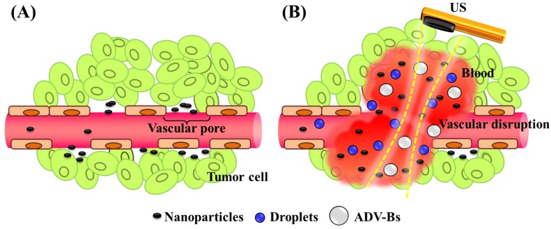
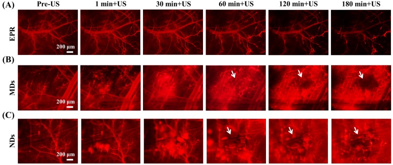
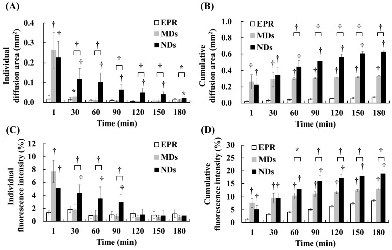
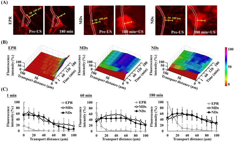
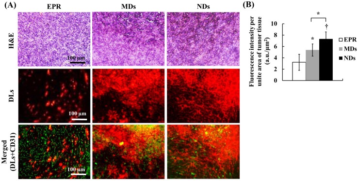
Drug penetration influences the efficacy of tumor therapy. Although the leaky vessels of tumors can improve the penetration of nanodrugs via the enhanced permeability and retention (EPR) effect, various aspects of the tumor microenvironment still restrict this process. This study investigated whether vascular disruption using the acoustic vaporization of micro- or nanoscale droplets (MDs or NDs) induced by ultrasound sonication can overcome the limitations of the EPR effect to allow drug diffusion into extensive regions. The intravital penetration of DiI-labeled liposomes (as a drug model with red fluorescence) was observed using an acousto-optical integrated system comprising a 2-MHz focused ultrasound transducer (transmitting a three-cycle single pulse and a peak negative pressure of 10 MPa) in a window-chamber mouse model. Histology images of the solid tumor were also used to quantify and demonstrate the locations where DiI-labeled liposomes accumulated. In the intravital image analyses, the cumulative diffusion area and fluorescence intensity at 180 min were 0.08±0.01 mm(2) (mean±standard deviation) and 8.5±0.4%, respectively, in the EPR group, 0.33±0.01 mm(2) and 13.1±0.4% in the MD group (p<0.01), and 0.63±0.01 mm(2) and 18.9±1.1% in the ND group (p<0.01). The intratumoral accumulations of DiI-labeled liposomes were 1.7- and 2.3-fold higher in the MD and ND groups, respectively, than in the EPR group. These results demonstrate that vascular disruption induced by acoustic droplet vaporization can improve drug penetration more than utilizing the EPR effect. The NDs showed longer lifetime in vivo than MDs and provided potential abilities of long periods of treatment, intertissue ND vaporization, and intertissue NDs-converted bubble cavitation to improve the drug penetration and transport distance.
药物渗透会影响肿瘤治疗的疗效。尽管肿瘤的渗漏血管可通过增强渗透与滞留(EPR)效应提高纳米药物的渗透,但肿瘤微环境的各个方面仍会限制这一过程。本研究调查了利用超声声处理诱导微米或纳米级液滴(MDs或NDs)的声致汽化造成血管破坏是否能够克服EPR效应的局限性,使药物扩散至更广泛区域。在窗室小鼠模型中,使用由一个2兆赫聚焦超声换能器(发射一个三周期单脉冲且峰值负压为10兆帕)组成的声光集成系统,观察DiI标记脂质体(作为具有红色荧光的药物模型)的活体渗透情况。实体瘤的组织学图像也用于量化并展示DiI标记脂质体积累的位置。在活体图像分析中,EPR组在180分钟时的累积扩散面积和荧光强度分别为0.08±0.01平方毫米(平均值±标准差)和8.5±0.4%,MD组为0.33±0.01平方毫米和13.1±0.4%(p<0.01),ND组为0.63±0.01平方毫米和18.9±1.1%(p<0.01)。DiI标记脂质体在MD组和ND组中的瘤内积累量分别比EPR组高1.7倍和2.3倍。这些结果表明,声致液滴汽化诱导的血管破坏比利用EPR效应能更好地改善药物渗透。NDs在体内的存留时间比MDs长,并具有长时间治疗、组织间ND汽化以及组织间NDs转化的气泡空化等潜在能力,可改善药物渗透和运输距离。