• 文献检索
  • 文档翻译
  • 深度研究
  • 学术资讯
  • Suppr Zotero 插件Zotero 插件
  • 邀请有礼
  • 套餐&价格
  • 历史记录
应用&插件
Suppr Zotero 插件Zotero 插件浏览器插件Mac 客户端Windows 客户端微信小程序
定价
高级版会员购买积分包购买API积分包
服务
文献检索文档翻译深度研究API 文档MCP 服务
关于我们
关于 Suppr公司介绍联系我们用户协议隐私条款
关注我们

Suppr 超能文献

核心技术专利:CN118964589B侵权必究
粤ICP备2023148730 号-1Suppr @ 2026

文献检索

告别复杂PubMed语法,用中文像聊天一样搜索,搜遍4000万医学文献。AI智能推荐,让科研检索更轻松。

立即免费搜索

文件翻译

保留排版,准确专业,支持PDF/Word/PPT等文件格式,支持 12+语言互译。

免费翻译文档

深度研究

AI帮你快速写综述,25分钟生成高质量综述,智能提取关键信息,辅助科研写作。

立即免费体验

基于超广角光学相干断层扫描的视网膜微血管造影成像技术

Wide-field optical coherence tomography based microangiography for retinal imaging.

作者信息

Zhang Qinqin, Lee Cecilia S, Chao Jennifer, Chen Chieh-Li, Zhang Thomas, Sharma Utkarsh, Zhang Anqi, Liu Jin, Rezaei Kasra, Pepple Kathryn L, Munsen Richard, Kinyoun James, Johnstone Murray, Van Gelder Russell N, Wang Ruikang K

机构信息

University of Washington, Department of Bioengineering, 3720 15th Ave NE, Seattle, WA 98195, USA.

University of Washington, Department of Ophthalmology, 325 Ninth Avenue, Seattle, WA 98104, USA.

出版信息

Sci Rep. 2016 Feb 25;6:22017. doi: 10.1038/srep22017.

DOI:10.1038/srep22017
PMID:26912261
原文链接:https://pmc.ncbi.nlm.nih.gov/articles/PMC4766473/
Abstract

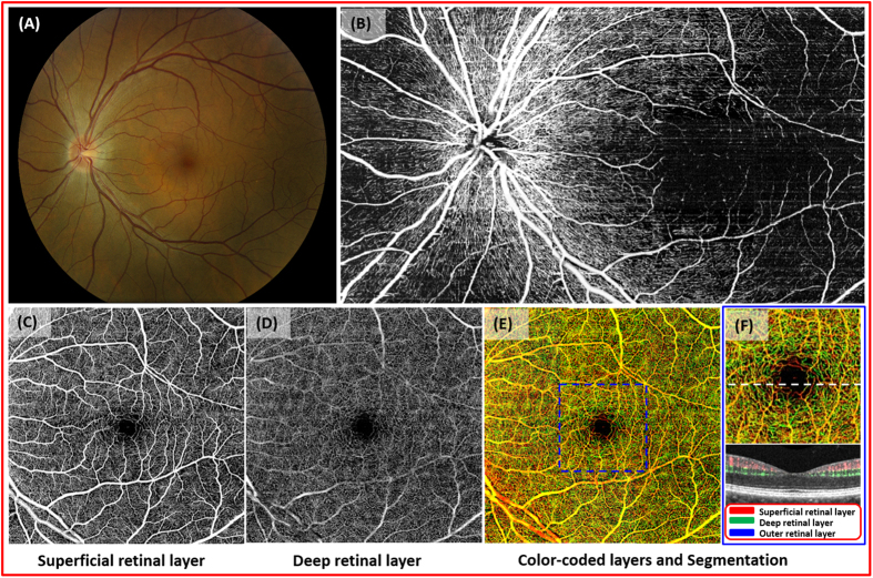
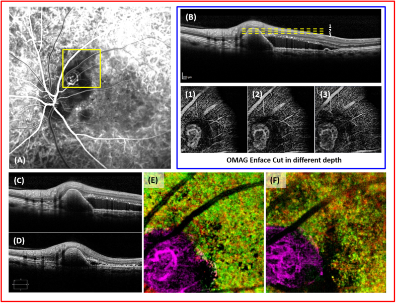
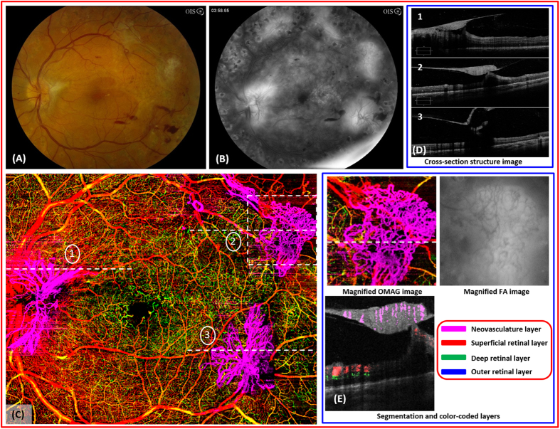
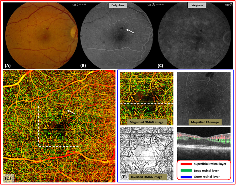
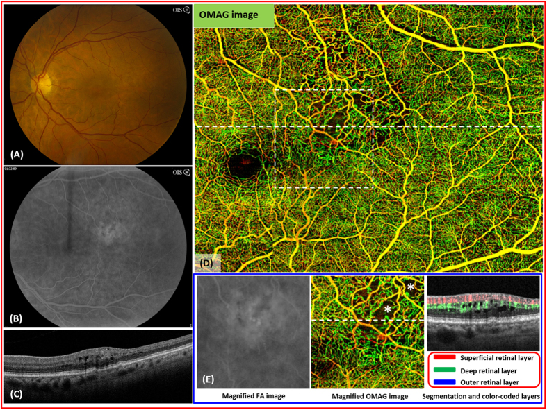
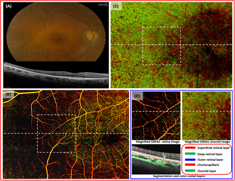
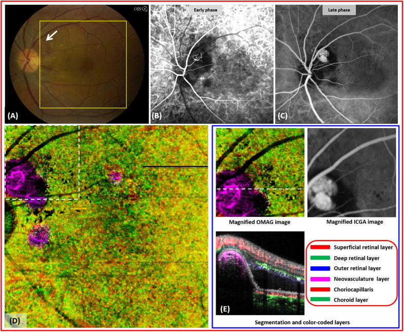
Optical coherence tomography angiography (OCTA) allows for the evaluation of functional retinal vascular networks without a need for contrast dyes. For sophisticated monitoring and diagnosis of retinal diseases, OCTA capable of providing wide-field and high definition images of retinal vasculature in a single image is desirable. We report OCTA with motion tracking through an auxiliary real-time line scan ophthalmoscope that is clinically feasible to image functional retinal vasculature in patients, with a coverage of more than 60 degrees of retina while still maintaining high definition and resolution. We demonstrate six illustrative cases with unprecedented details of vascular involvement in retinal diseases. In each case, OCTA yields images of the normal and diseased microvasculature at all levels of the retina, with higher resolution than observed with fluorescein angiography. Wide-field OCTA technology will be an important next step in augmenting the utility of OCT technology in clinical practice.

摘要

光学相干断层扫描血管造影(OCTA)无需使用造影剂就能评估视网膜功能血管网络。对于视网膜疾病的精密监测和诊断,能够在单幅图像中提供视网膜血管系统的宽视野和高清晰度图像的OCTA是理想之选。我们报告了一种通过辅助实时线扫描检眼镜进行运动跟踪的OCTA,它在临床上对患者功能性视网膜血管成像切实可行,可覆盖超过60度的视网膜,同时仍能保持高清晰度和分辨率。我们展示了六个具有前所未有的视网膜疾病血管受累细节的典型病例。在每个病例中,OCTA都能生成视网膜各层正常和病变微血管的图像,分辨率高于荧光素血管造影。宽视野OCTA技术将是增强OCT技术在临床实践中效用的重要下一步。

https://cdn.ncbi.nlm.nih.gov/pmc/blobs/c5b0/4766473/a481ee1ec32f/srep22017-f7.jpg
https://cdn.ncbi.nlm.nih.gov/pmc/blobs/c5b0/4766473/064ab28d7db2/srep22017-f1.jpg
https://cdn.ncbi.nlm.nih.gov/pmc/blobs/c5b0/4766473/7a36a56c66ff/srep22017-f2.jpg
https://cdn.ncbi.nlm.nih.gov/pmc/blobs/c5b0/4766473/823d792adbbf/srep22017-f3.jpg
https://cdn.ncbi.nlm.nih.gov/pmc/blobs/c5b0/4766473/2e6f728a6ba2/srep22017-f4.jpg
https://cdn.ncbi.nlm.nih.gov/pmc/blobs/c5b0/4766473/0dbfa38fe449/srep22017-f5.jpg
https://cdn.ncbi.nlm.nih.gov/pmc/blobs/c5b0/4766473/0cf95ee1a3ed/srep22017-f6.jpg
https://cdn.ncbi.nlm.nih.gov/pmc/blobs/c5b0/4766473/a481ee1ec32f/srep22017-f7.jpg

相似文献

1
Wide-field optical coherence tomography based microangiography for retinal imaging.基于超广角光学相干断层扫描的视网膜微血管造影成像技术
Sci Rep. 2016 Feb 25;6:22017. doi: 10.1038/srep22017.
2
Retinal and choroidal vascular features in patients with retinitis pigmentosa imaged by OCT based microangiography.基于光学相干断层扫描血管造影成像的色素性视网膜炎患者的视网膜和脉络膜血管特征
Graefes Arch Clin Exp Ophthalmol. 2017 Jul;255(7):1287-1295. doi: 10.1007/s00417-017-3633-x. Epub 2017 Mar 17.
3
Select Features of Diabetic Retinopathy on Swept-Source Optical Coherence Tomographic Angiography Compared With Fluorescein Angiography and Normal Eyes.比较扫频源光学相干断层血管造影与荧光素血管造影和正常眼的糖尿病视网膜病变特征。
JAMA Ophthalmol. 2016 Jun 1;134(6):644-50. doi: 10.1001/jamaophthalmol.2016.0600.
4
Visualization of the Retinal Vasculature Using Wide-Field Montage Optical Coherence Tomography Angiography.使用广角蒙太奇光学相干断层扫描血管造影术可视化视网膜血管系统
Ophthalmic Surg Lasers Imaging Retina. 2015 Jun;46(6):611-6. doi: 10.3928/23258160-20150610-03.
5
The application of optical coherence tomography angiography in retinal diseases.光学相干断层扫描血管造影在视网膜疾病中的应用。
Surv Ophthalmol. 2017 Nov-Dec;62(6):838-866. doi: 10.1016/j.survophthal.2017.05.006. Epub 2017 Jun 1.
6
High-definition and 3-dimensional imaging of macular pathologies with high-speed ultrahigh-resolution optical coherence tomography.利用高速超高分辨率光学相干断层扫描技术对黄斑病变进行高清三维成像。
Ophthalmology. 2006 Nov;113(11):2054.e1-14. doi: 10.1016/j.ophtha.2006.05.046.
7
Optical Coherence Tomography Angiography of Diabetic Retinopathy in Human Subjects.人体糖尿病视网膜病变的光学相干断层扫描血管造影
Ophthalmic Surg Lasers Imaging Retina. 2015 Sep;46(8):796-805. doi: 10.3928/23258160-20150909-03.
8
Wide-field en face swept-source optical coherence tomography angiography using extended field imaging in diabetic retinopathy.宽视野正面对扫激光相干断层扫描血管造影术在糖尿病视网膜病变中的应用。
Br J Ophthalmol. 2018 Sep;102(9):1199-1203. doi: 10.1136/bjophthalmol-2017-311358. Epub 2017 Nov 29.
9
Optical Coherence Tomography Angiography of Chorioretinal Diseases.脉络膜视网膜疾病的光学相干断层扫描血管造影
Ophthalmic Surg Lasers Imaging Retina. 2016 Sep 1;47(9):848-61. doi: 10.3928/23258160-20160901-09.
10
Optical coherence tomography angiography: A comprehensive review of current methods and clinical applications.光学相干断层扫描血管造影:当前方法与临床应用的全面综述。
Prog Retin Eye Res. 2017 Sep;60:66-100. doi: 10.1016/j.preteyeres.2017.07.002. Epub 2017 Jul 29.

引用本文的文献

1
Widefield OCT angiography.广角光学相干断层扫描血管造影术
Prog Retin Eye Res. 2025 Jul;107:101378. doi: 10.1016/j.preteyeres.2025.101378. Epub 2025 Jun 13.
2
Advances in OCT Angiography.光学相干断层扫描血管造影术的进展。
Transl Vis Sci Technol. 2025 Mar 3;14(3):6. doi: 10.1167/tvst.14.3.6.
3
Wearable optical coherence tomography angiography probe with extended depth of field.具有扩展景深的可穿戴光学相干断层扫描血管造影探头。

本文引用的文献

1
Minimizing projection artifacts for accurate presentation of choroidal neovascularization in OCT micro-angiography.在光学相干断层扫描微血管造影中最小化投影伪影以准确呈现脉络膜新生血管。
Biomed Opt Express. 2015 Sep 28;6(10):4130-43. doi: 10.1364/BOE.6.004130. eCollection 2015 Oct 1.
2
Automated choroidal neovascularization detection algorithm for optical coherence tomography angiography.用于光学相干断层扫描血管造影的自动脉络膜新生血管检测算法
Biomed Opt Express. 2015 Aug 25;6(9):3564-76. doi: 10.1364/BOE.6.003564. eCollection 2015 Sep 1.
3
Wide-field imaging of retinal vasculature using optical coherence tomography-based microangiography provided by motion tracking.
J Biomed Opt. 2025 Jan;30(1):016003. doi: 10.1117/1.JBO.30.1.016003. Epub 2025 Jan 22.
4
Insight into the Brain: Application of the Retinal Microvasculature as a Biomarker for Cerebrovascular Diseases through Optical Coherence Tomography Angiography.洞察大脑:通过光学相干断层扫描血管造影术将视网膜微血管系统用作脑血管疾病生物标志物的应用
Curr Ophthalmol Rep. 2024 Mar;12(1):1-11. doi: 10.1007/s40135-023-00320-z. Epub 2023 Nov 28.
5
Tail Artifact Removal via Transmittance Effect Subtraction in Optical Coherence Tail Artifact Images.基于透过率效应差减的光学相干尾影伪像图像中尾影去除。
Sensors (Basel). 2023 Nov 21;23(23):9312. doi: 10.3390/s23239312.
6
Association of Male Sex and Microvascular Alterations on Optical Coherence Tomography Angiography in Diabetes.男性性别与糖尿病光学相干断层扫描血管造影微血管改变的相关性。
Transl Vis Sci Technol. 2023 Nov 1;12(11):30. doi: 10.1167/tvst.12.11.30.
7
Extended and adjustable field-of-view of variable interscan time analysis by ammonite-scanning swept-source optical coherence tomography angiography.通过菊石扫描扫频源光学相干断层扫描血管造影术进行可变帧间时间分析时的扩展和可调视野。
Biomed Opt Express. 2023 Jul 14;14(8):4112-4125. doi: 10.1364/BOE.491611. eCollection 2023 Aug 1.
8
Spotlight on Lattice Degeneration Imaging Techniques.聚焦于格子样变性成像技术。
Clin Ophthalmol. 2023 Aug 16;17:2383-2395. doi: 10.2147/OPTH.S405200. eCollection 2023.
9
Prospective clinical study of retinal microvascular alteration after ICL implantation.ICL植入术后视网膜微血管改变的前瞻性临床研究
Front Cell Dev Biol. 2023 Jan 19;11:1115822. doi: 10.3389/fcell.2023.1115822. eCollection 2023.
10
Multimodal Imaging, Tele-Education, and Telemedicine in Retinopathy of Prematurity.多模态成像、远程医学教育和早产儿视网膜病变。
Middle East Afr J Ophthalmol. 2022 Nov 23;29(1):38-50. doi: 10.4103/meajo.meajo_56_22. eCollection 2022 Jan-Mar.
利用基于光学相干断层扫描的微血管造影术通过运动追踪对视网膜血管系统进行广角成像。
J Biomed Opt. 2015 Jun;20(6):066008. doi: 10.1117/1.JBO.20.6.066008.
4
Advanced imaging for glaucoma study: design, baseline characteristics, and inter-site comparison.青光眼研究的高级成像:设计、基线特征和站点间比较。
Am J Ophthalmol. 2015 Feb;159(2):393-403.e2. doi: 10.1016/j.ajo.2014.11.010. Epub 2014 Nov 8.
5
Retinal vascular layers imaged by fluorescein angiography and optical coherence tomography angiography.荧光素血管造影和光相干断层扫描血管造影成像的视网膜血管层。
JAMA Ophthalmol. 2015 Jan;133(1):45-50. doi: 10.1001/jamaophthalmol.2014.3616.
6
Swept-source OCT angiography of macular telangiectasia type 2.2型黄斑毛细血管扩张症的扫频源光学相干断层扫描血管造影
Ophthalmic Surg Lasers Imaging Retina. 2014 Sep-Oct;45(5):369-80. doi: 10.3928/23258160-20140909-06.
7
Reevaluating the definition of intraretinal microvascular abnormalities and neovascularization elsewhere in diabetic retinopathy using optical coherence tomography and fluorescein angiography.利用光学相干断层扫描和荧光素血管造影术重新评估糖尿病视网膜病变中视网膜内微血管异常和其他部位新生血管形成的定义。
Am J Ophthalmol. 2015 Jan;159(1):101-10.e1. doi: 10.1016/j.ajo.2014.09.041. Epub 2014 Oct 25.
8
Swept-source OCT angiography of the retinal vasculature using intensity differentiation-based optical microangiography algorithms.使用基于强度分化的光学微血管造影算法对视网膜血管系统进行扫频源光学相干断层扫描血管造影。
Ophthalmic Surg Lasers Imaging Retina. 2014 Sep-Oct;45(5):382-9. doi: 10.3928/23258160-20140909-08.
9
User-guided segmentation for volumetric retinal optical coherence tomography images.用于视网膜光学相干断层扫描容积图像的用户引导分割
J Biomed Opt. 2014 Aug;19(8):086020. doi: 10.1117/1.JBO.19.8.086020.
10
Recent advances of stem cell therapy for retinitis pigmentosa.视网膜色素变性干细胞治疗的最新进展
Int J Mol Sci. 2014 Aug 20;15(8):14456-74. doi: 10.3390/ijms150814456.