Weir-McCall Jonathan R, Duce Suzanne L, Gandy Stephen J, Matthew Shona Z, Martin Patricia, Cassidy Deirdre B, McCormick Lynne, Belch Jill J F, Struthers Allan D, Colhoun Helen M, Houston J Graeme
Division of Cardiovascular and Diabetes Medicine, Medical Research Institute, University of Dundee, ᅟ, DD1 9SY, UK.
NHS Tayside Clinical Radiology, Ninewells Hospital, Dundee, DD1 9SY, UK.
BMC Med Imaging. 2016 Feb 29;16:18. doi: 10.1186/s12880-016-0121-4.
The aim of this study was to use whole body cardiovascular magnetic resonance imaging (WB CVMR) to assess the heart and arterial network in a single examination, so as to describe the burden of atherosclerosis and subclinical disease in participants with symptomatic single site vascular disease.
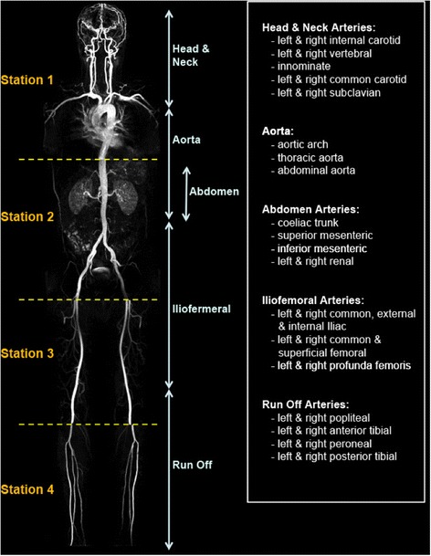
64 patients with a history of symptomatic single site vascular disease (38 coronary artery disease (CAD), 9 cerebrovascular disease, 17 peripheral arterial disease (PAD)) underwent whole body angiogram and cardiac MR in a 3 T scanner. The arterial tree was subdivided into 31 segments and each scored according to the degree of stenosis. From this a standardised atheroma score (SAS) was calculated. Cine and late gadolinium enhancement images of the left ventricle were obtained.
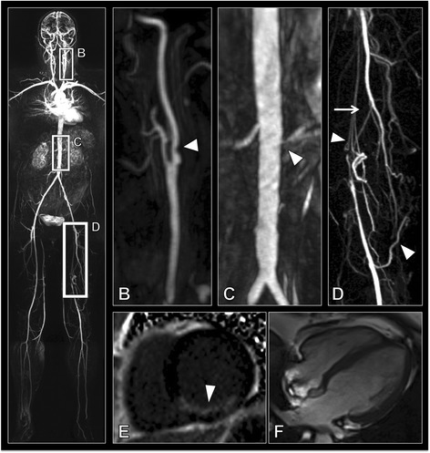
Asymptomatic atherosclerotic disease with greater than 50% stenosis in arteries other than that responsible for their presenting complain was detected in 37% of CAD, 33% of cerebrovascular and 47% of PAD patients. Unrecognised myocardial infarcts were observed in 29% of PAD patients. SAS was significantly higher in PAD patients 24 (17.5-30.5) compared to CAD 4 (2-11.25) or cerebrovascular disease patients 6 (2-10) (ANCOVA p < 0.001). Standardised atheroma score positively correlated with age (β 0.36 p = 0.002), smoking status (β 0.34 p = 0.002), and LV mass (β -0.61 p = 0.001) on multiple linear regression.
WB CVMR is an effective method for the stratification of cardiovascular disease. The high prevalence of asymptomatic arterial disease, and silent myocardial infarctions, particularly in the peripheral arterial disease group, demonstrates the importance of a systematic approach to the assessment of cardiovascular disease.
本研究的目的是利用全身心血管磁共振成像(WB CVMR)在一次检查中评估心脏和动脉网络,以描述有症状的单部位血管疾病参与者的动脉粥样硬化和亚临床疾病负担。
64例有症状的单部位血管疾病患者(38例冠状动脉疾病(CAD)、9例脑血管疾病、17例外周动脉疾病(PAD))在3T扫描仪上进行了全身血管造影和心脏磁共振成像。动脉树被细分为31个节段,并根据狭窄程度进行评分。由此计算出标准化动脉粥样硬化评分(SAS)。获取了左心室的电影图像和延迟钆增强图像。
在37%的CAD患者、33%的脑血管疾病患者和47%的PAD患者中检测到除导致其当前症状的动脉外,其他动脉存在狭窄程度大于50%的无症状动脉粥样硬化疾病。在29%的PAD患者中观察到未被识别的心肌梗死。与CAD患者的4(2 - 11.25)或脑血管疾病患者的6(2 - 10)相比,PAD患者的SAS显著更高(24(17.5 - 30.5))(协方差分析p < 0.001)。在多元线性回归中,标准化动脉粥样硬化评分与年龄(β 0.36 p = 0.002)、吸烟状况(β 0.34 p = 0.002)和左心室质量(β -0.61 p = 0.001)呈正相关。
WB CVMR是一种有效的心血管疾病分层方法。无症状动脉疾病和无症状心肌梗死的高患病率,特别是在外周动脉疾病组中,表明了系统评估心血管疾病方法的重要性。