Rasta Seyed Hossein, Nikfarjam Shima, Javadzadeh Alireza
Department of Medical Bioengineering, Stem Cell Research Center, Tabriz University of Medical Sciences, Tabriz, Iran ; School of Medical Sciences, University of Aberdeen, Aberdeen, UK.
Department of Medical Bioengineering, Stem Cell Research Center, Tabriz University of Medical Sciences, Tabriz, Iran.
Bioimpacts. 2015;5(4):183-90. doi: 10.15171/bi.2015.27. Epub 2015 Dec 28.
Retinal capillary nonperfusion (CNP) is one of the retinal vascular diseases in diabetic retinopathy (DR) patients. As there is no comprehensive detection technique to recognize CNP areas, we proposed a different method for computing detection of ischemic retina, non-perfused (NP) regions, in fundus fluorescein angiogram (FFA) images.
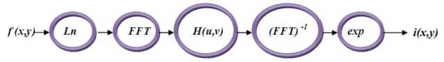
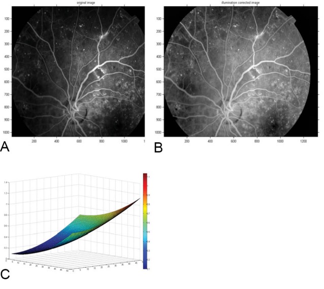
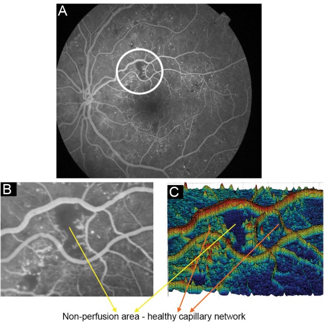
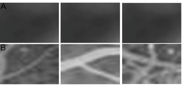
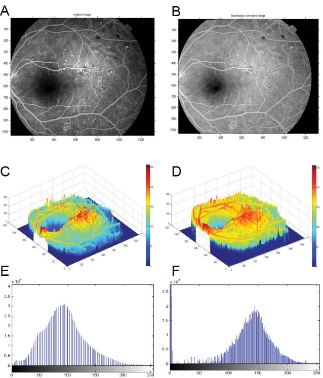
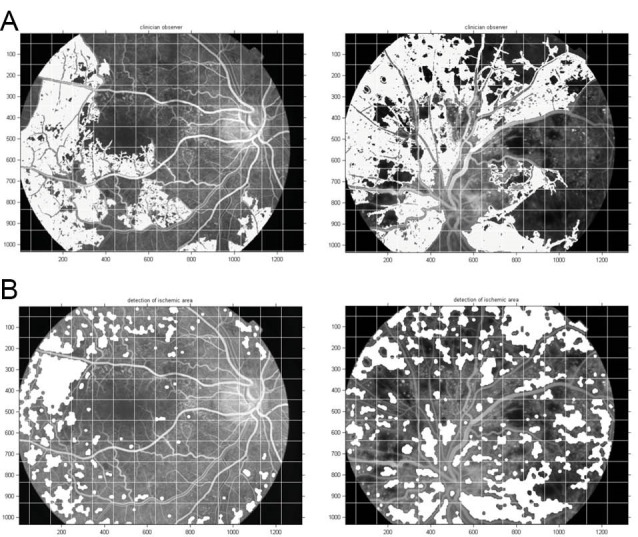
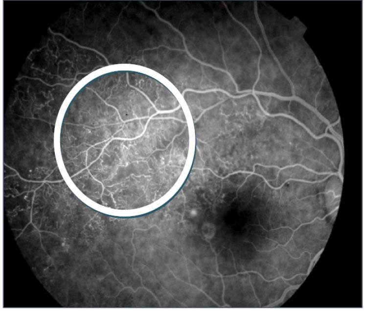
Whilst major vessels appear as ridges, non-perfused areas are usually observed as ponds that are surrounded by healthy capillaries in FFA images. A new technique using homomorphic filtering to correct light illumination and detect the ponds surrounded in healthy capillaries on FFA images was designed and applied on DR fundus images. These images were acquired from the diabetic patients who had referred to the Nikookari hospital and were diagnosed for diabetic retinopathy during one year. Our strategy was screening the whole image with a fixed window size, which is small enough to enclose areas with identified topographic characteristics. To discard false nominees, we also performed a thresholding operation on the screen and marked images. To validate its performance we applied our detection algorithm on 41 FFA diabetic retinopathy fundus images in which the CNP areas were manually delineated by three clinical experts.
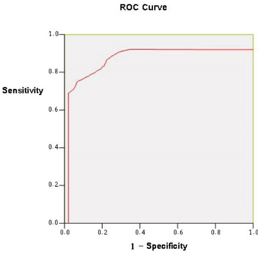
Lesions were found as smooth regions with very high uniformity, low entropy, and small intensity variations in FFA images. The results of automated detection method were compared with manually marked CNP areas so achieved sensitivity of 81%, specificity of 78%, and accuracy of 91%.The result was present as a Receiver operating character (ROC) curve, which has an area under the curve (AUC) of 0.796 with 95% confidence intervals.
This technique introduced a new automated detection algorithm to recognize non-perfusion lesions on FFA. This has potential to assist detecting and managing of ischemic retina and may be incorporated into automated grading diabetic retinopathy structures.
视网膜毛细血管无灌注(CNP)是糖尿病视网膜病变(DR)患者的视网膜血管疾病之一。由于目前尚无全面的检测技术来识别CNP区域,我们提出了一种不同的方法来计算眼底荧光血管造影(FFA)图像中缺血视网膜、无灌注(NP)区域的检测。
在FFA图像中,主要血管呈现为脊状,而无灌注区域通常表现为被健康毛细血管包围的池状。设计了一种使用同态滤波来校正光照并检测FFA图像中被健康毛细血管包围的池状区域的新技术,并将其应用于DR眼底图像。这些图像来自转诊至尼库卡里医院并在一年内被诊断为糖尿病视网膜病变的糖尿病患者。我们的策略是使用固定窗口大小对整个图像进行筛查,该窗口大小要足够小,以包围具有特定地形特征的区域。为了排除错误的候选区域,我们还对筛选后的图像进行了阈值操作并进行标记。为了验证其性能,我们将检测算法应用于41张FFA糖尿病视网膜病变眼底图像,其中三位临床专家手动勾勒出了CNP区域。
在FFA图像中,病变表现为均匀性非常高、熵低且强度变化小的平滑区域。将自动检测方法的结果与手动标记的CNP区域进行比较,灵敏度达到81%,特异性为78%,准确率为91%。结果以受试者工作特征(ROC)曲线呈现,曲线下面积(AUC)为0.796,95%置信区间。
该技术引入了一种新的自动检测算法,用于识别FFA上的无灌注病变。这有可能辅助检测和管理缺血性视网膜,并且可能被纳入糖尿病视网膜病变结构的自动分级中。