Kiselinova Maja, De Spiegelaere Ward, Buzon Maria Jose, Malatinkova Eva, Lichterfeld Mathias, Vandekerckhove Linos
HIV Translational Research Unit (HTRU), Department of Internal Medicine, Ghent University and Ghent University Hospital, Ghent, Belgium.
Ragon Institute of MGH, MIT and Harvard, Boston, Massachusetts, United States of America.
PLoS Pathog. 2016 Mar 3;12(3):e1005472. doi: 10.1371/journal.ppat.1005472. eCollection 2016 Mar.
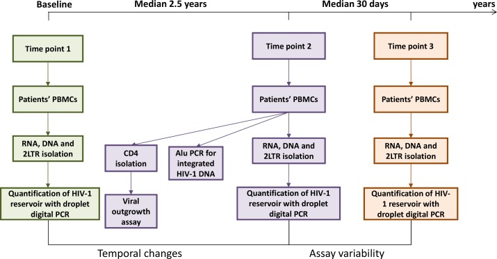
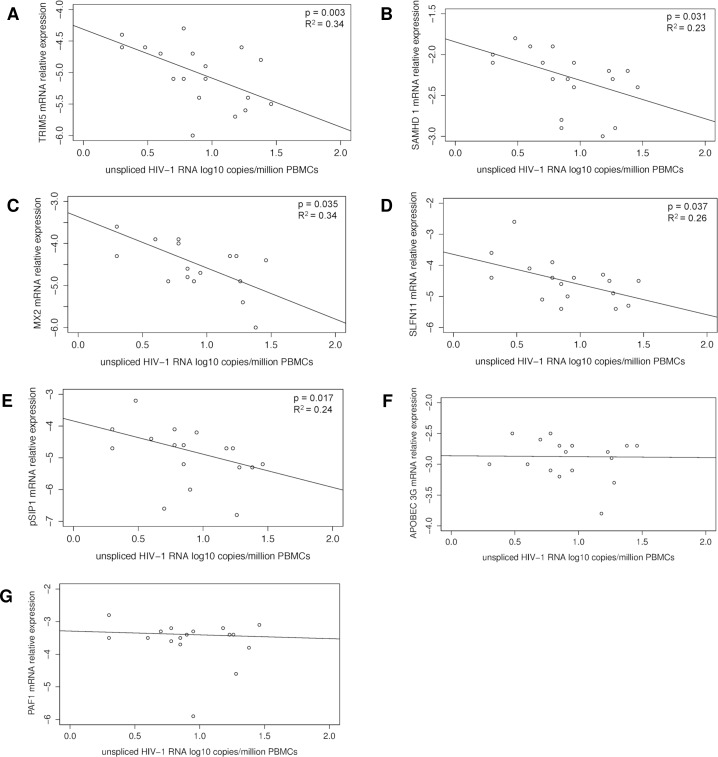
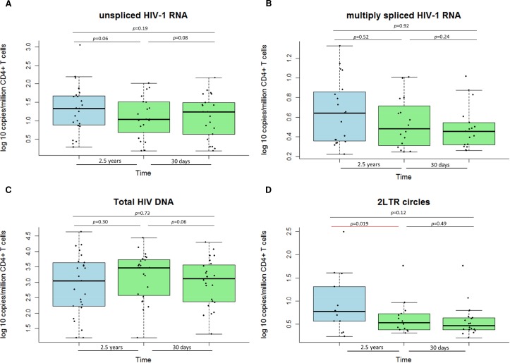
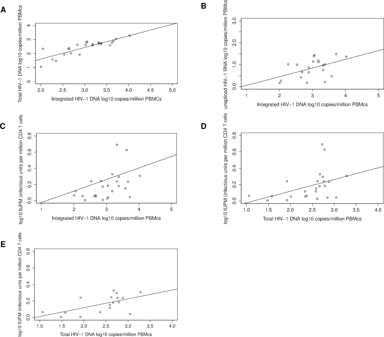
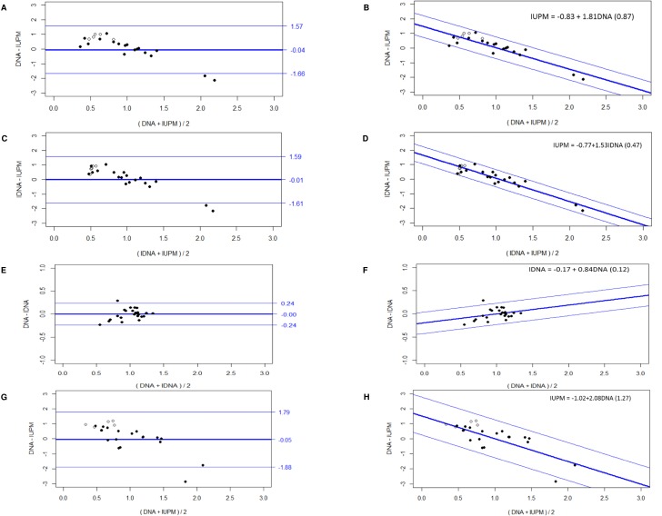
The persistence of a reservoir of latently infected CD4 T cells remains one of the major obstacles to cure HIV. Numerous strategies are being explored to eliminate this reservoir. To translate these efforts into clinical trials, there is a strong need for validated biomarkers that can monitor the reservoir over time in vivo. A comprehensive study was designed to evaluate and compare potential HIV-1 reservoir biomarkers. A cohort of 25 patients, treated with suppressive antiretroviral therapy was sampled at three time points, with median of 2.5 years (IQR: 2.4-2.6) between time point 1 and 2; and median of 31 days (IQR: 28-36) between time point 2 and 3. Patients were median of 6 years (IQR: 3-12) on ART, and plasma viral load (<50 copies/ml) was suppressed for median of 4 years (IQR: 2-8). Total HIV-1 DNA, unspliced (us) and multiply spliced HIV-1 RNA, and 2LTR circles were quantified by digital PCR in peripheral blood, at 3 time points. At the second time point, a viral outgrowth assay (VOA) was performed, and integrated HIV-1 DNA and relative mRNA expression levels of HIV-1 restriction factors were quantified. No significant change was found for long- and short-term dynamics of all HIV-1 markers tested in peripheral blood. Integrated HIV-1 DNA was associated with total HIV-1 DNA (p<0.001, R² = 0.85), us HIV-1 RNA (p = 0.029, R² = 0.40), and VOA (p = 0.041, R2 = 0.44). Replication-competent virus was detected in 80% of patients by the VOA and it correlated with total HIV-1 DNA (p = 0.039, R² = 0.54). The mean quantification difference between Alu-PCR and VOA was 2.88 log10, and 2.23 log10 between total HIV-1 DNA and VOA. The levels of usHIV-1 RNA were inversely correlated with mRNA levels of several HIV-1 restriction factors (TRIM5α, SAMHD1, MX2, SLFN11, pSIP1). Our study reveals important correlations between the viral outgrowth and total and integrated HIV-1 DNA measures, suggesting that the total pool of HIV-1 DNA may predict the size of the replication-competent virus in ART suppressed patients.
潜伏感染的CD4 T细胞库的持续存在仍然是治愈HIV的主要障碍之一。目前正在探索多种策略来消除这个细胞库。为了将这些努力转化为临床试验,迫切需要能够在体内长期监测该细胞库的经过验证的生物标志物。一项全面的研究旨在评估和比较潜在的HIV-1细胞库生物标志物。对25名接受抑制性抗逆转录病毒治疗的患者进行队列研究,在三个时间点进行采样,时间点1和2之间的中位数为2.5年(四分位间距:2.4 - 2.6);时间点2和3之间的中位数为31天(四分位间距:28 - 36)。患者接受抗逆转录病毒治疗的中位数为6年(四分位间距:3 - 12),血浆病毒载量(<50拷贝/ml)被抑制的中位数为4年(四分位间距:2 - 8)。在三个时间点通过数字PCR对外周血中的总HIV-1 DNA、未剪接(us)和多重剪接的HIV-1 RNA以及2LTR环进行定量。在第二个时间点,进行病毒增殖试验(VOA),并对整合的HIV-1 DNA和HIV-1限制因子的相对mRNA表达水平进行定量。在所检测的外周血中所有HIV-1标志物的长期和短期动态变化方面未发现显著变化。整合的HIV-1 DNA与总HIV-1 DNA(p<0.001,R² = 0.85)、us HIV-1 RNA(p = 0.029,R² = 0.40)以及VOA(p = 0.041,R2 = 0.44)相关。通过VOA在80%的患者中检测到具有复制能力的病毒,并且它与总HIV-1 DNA相关(p = 0.039,R² = 0.54)。Alu-PCR和VOA之间的平均定量差异为2.88 log10,总HIV-1 DNA和VOA之间为2.23 log10。usHIV-1 RNA的水平与几种HIV-1限制因子(TRIM5α、SAMHD1、MX2、SLFN11、pSIP1)的mRNA水平呈负相关。我们的研究揭示了病毒增殖与总HIV-1 DNA和整合HIV-1 DNA测量值之间的重要相关性,表明HIV-1 DNA的总量可能预测抗逆转录病毒治疗抑制患者中具有复制能力的病毒的大小。