Rashidian Mohammad, Keliher Edmund, Dougan Michael, Juras Patrick K, Cavallari Marco, Wojtkiewicz Gregory R, Jacobsen Johanne, Edens Jerre G, Tas Jeroen M G, Victora Gabriel, Weissleder Ralph, Ploegh Hidde
Whitehead Institute for Biomedical Research, Cambridge, MA 02142; Department of Biology, Massachusetts Institute of Technology, Cambridge, MA 02142.
Center for Systems Biology Department, Massachusetts General Hospital, 185 Cambridge St., Boston, MA 02114 (USA).
ACS Cent Sci. 2015 Jun 24;1(3):142-147. doi: 10.1021/acscentsci.5b00121. Epub 2015 Jun 3.
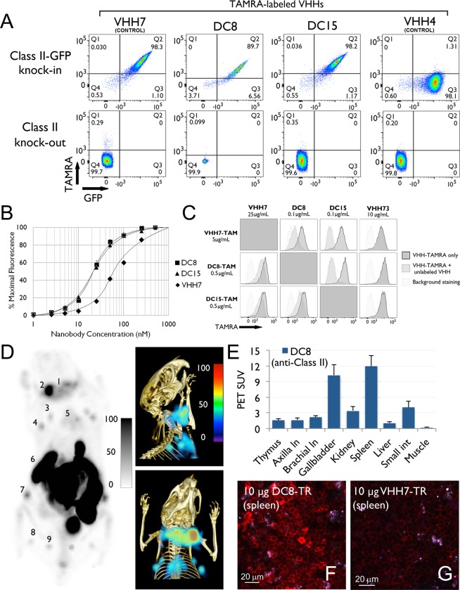
We generated F-labeled antibody fragments for PET imaging using a sortase-mediated reaction to install a transcyclooctene (TCO)-functionalized short peptide onto proteins of interest, followed by reaction with a tetrazine-labeled-F-2-deoxyfluoroglucose (FDG). The method is rapid, robust, and site-specific (radiochemical yields >25%, not decay corrected). The availability of F-2-deoxyfluoroglucose avoids the need for more complicated chemistries used to generate carbon-fluorine bonds. We demonstrate the utility of the method by detecting heterotopic pancreatic tumors in mice by PET, using anti-Class II MHC single domain antibodies. We correlate macroscopic PET images with microscopic two-photon visualization of the tumor. Our approach provides easy access to F-labeled antibodies and their fragments at a level of molecular specificity that complements conventionalF-FDG imaging.
我们使用分选酶介导的反应生成用于正电子发射断层扫描(PET)成像的F标记抗体片段,即将反式环辛烯(TCO)功能化的短肽安装到感兴趣的蛋白质上,然后与四嗪标记的F-2-脱氧氟葡萄糖(FDG)反应。该方法快速、稳健且具有位点特异性(放射化学产率>25%,未进行衰变校正)。F-2-脱氧氟葡萄糖的可用性避免了使用更复杂的化学方法来生成碳-氟键的需求。我们通过使用抗II类主要组织相容性复合体(MHC)单域抗体,在小鼠中通过PET检测异位胰腺肿瘤来证明该方法的实用性。我们将宏观PET图像与肿瘤的微观双光子可视化相关联。我们的方法以补充传统F-FDG成像的分子特异性水平,提供了轻松获得F标记抗体及其片段的途径。