Department of Radiology , Duke University Medical Center , Durham , North Carolina 27710 , United States.
In vivo Cellular and Molecular Imaging laboratory , Vrije Universiteit Brussel , 1090 , Brussels , Belgium.
Bioconjug Chem. 2018 Dec 19;29(12):4090-4103. doi: 10.1021/acs.bioconjchem.8b00699. Epub 2018 Nov 14.
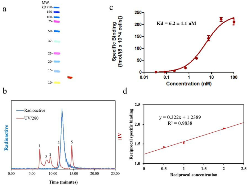
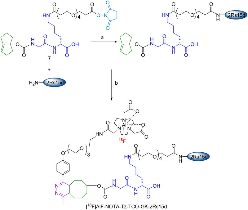
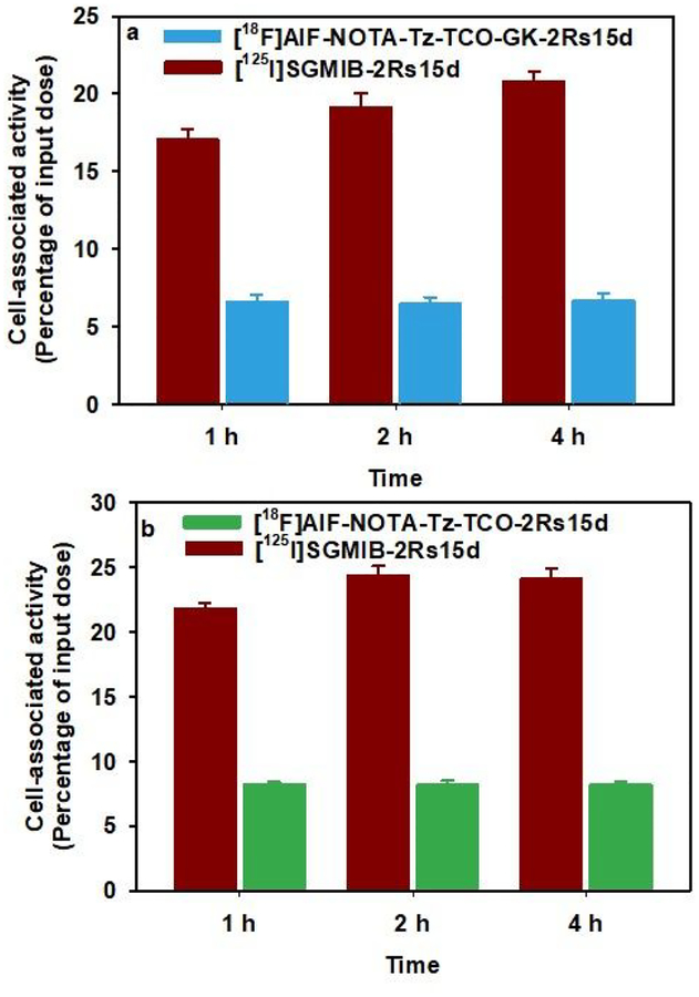
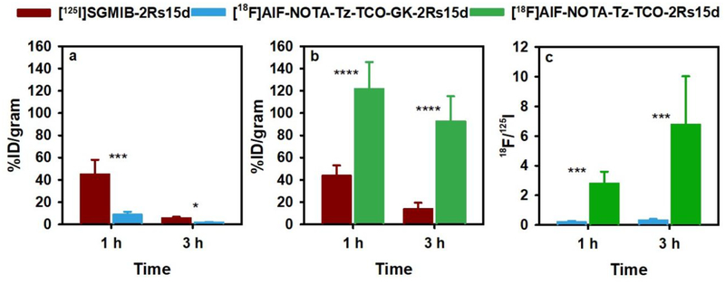
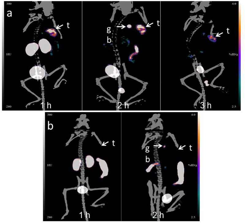
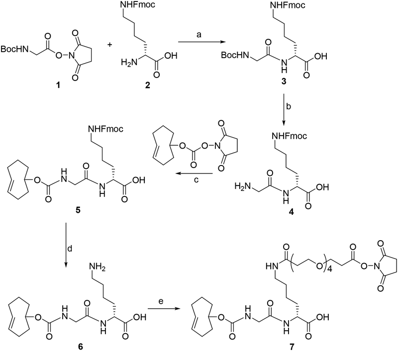
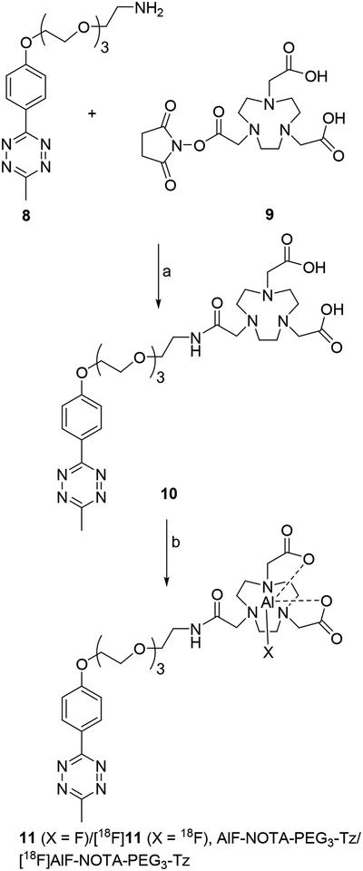
Single domain antibody fragments (sdAbs) labeled with F have shown promise for assessing the status of oncological targets such as the human epidermal growth factor receptor 2 (HER2) by positron emission tomography (PET). Earlier, we evaluated two residualizing prosthetic agents for F-labeling of anti-HER2 sdAbs; however, these methods resulted in poor labeling yields and high uptake of F activity in the kidneys. To potentially mitigate these limitations, we have now developed an F labeling method that utilizes the trans-cyclooctene (TCO)-tetrazine (Tz)-based inverse-electron demand Diels-Alder reaction (IEDDAR) in tandem with a renal brush border enzyme-cleavable glycine-lysine (GK) linker in the prosthetic moiety. The HER2-targeted sdAb 2Rs15d was derivatized with TCO-GK-PEG-NHS or TCO-PEG-NHS, which lacks the cleavable linker. As an additional control, the non HER2-specific sdAb R3B23 was derivatized with TCO-GK-PEG-NHS. The resultant sdAb conjugates were labeled with F by IEDDAR using [F]AlF-NOTA-PEG-methyltetrazine. As a positive control, the 2Rs15d sdAb was radioiodinated using the well-characterized residualizing prosthetic agent, N-succinimidyl 4-guanidinomethyl-3-[I]iodobenzoate ([I]SGMIB). Synthesis of [F]AlF-NOTA-Tz-TCO-GK-2Rs15d was achieved with an overall radiochemical yield (RCY) of 17.8 ± 1.5% ( n = 5) in 90 min, a significant improvement over prior methods (3-4% in 2-3 h). In vitro assays indicated that [F]AlF-NOTA-Tz-TCO-GK-2Rs15d bound with high affinity and immunoreactivity to HER2. In normal mice, when normalized to coinjected [I]SGMIB-2Rs15d, the kidney uptake of [F]AlF-NOTA-Tz-TCO-GK-2Rs15d was 15- and 28-fold lower ( P < 0.001) than that seen for the noncleavable control ([F]AlF-NOTA-Tz-TCO-2Rs15d) at 1 and 3 h, respectively. Uptake of [F]AlF-NOTA-Tz-TCO-GK-2Rs15d in HER2-expressing SKOV-3 ovarian carcinoma xenografts implanted in athymic mice was about 80% of that seen for coinjected [I]SGMIB-2Rs15d. On the other hand, kidney uptake was 5-6-fold lower, and as a result, tumor-to-kidney ratios were 4-fold higher for [F]AlF-NOTA-Tz-TCO-GK-2Rs15d than those for [I]SGMIB-2Rs15d. SKOV-3 xenografts were clearly delineated even at 1 h after administration of [F]AlF-NOTA-Tz-TCO-GK-2Rs15d by Micro-PET/CT imaging with even higher contrast observed thereafter. In conclusion, this strategy warrants further evaluation for labeling small proteins such as sdAbs because it offers the benefits of good radiochemical yields and enhanced tumor-to-normal tissue ratios, particularly in the kidney.
单域抗体片段(sdAbs)与 F 标记物结合,通过正电子发射断层扫描(PET)显示出评估人类表皮生长因子受体 2(HER2)等肿瘤靶标状态的潜力。早些时候,我们评估了两种用于抗 HER2 sdAb 的残留化 prosthetic 试剂进行 F 标记;然而,这些方法导致标记产率差,并且 F 活性在肾脏中的摄取量高。为了潜在地减轻这些限制,我们现在开发了一种 F 标记方法,该方法利用反电子需求 Diels-Alder 反应(IEDDAR)与 prosthetic 部分中的肾脏刷状缘酶可切割的甘氨酸-赖氨酸(GK)接头串联使用。靶向 HER2 的 sdAb 2Rs15d 用 TCO-GK-PEG-NHS 或 TCO-PEG-NHS 衍生化,后者缺乏可切割的接头。作为附加对照,非 HER2 特异性 sdAb R3B23 用 TCO-GK-PEG-NHS 衍生化。所得 sdAb 缀合物通过 IEDDAR 用 [F]AlF-NOTA-PEG-甲基四嗪(Tz-TCO-GK-PEG-NHS)进行 F 标记。作为阳性对照,2Rs15d sdAb 使用经过充分表征的残留化 prosthetic 试剂 N-琥珀酰亚胺基 4-胍基甲基-3-[I]碘代苯甲酸酯([I]SGMIB)进行放射性碘标记。[F]AlF-NOTA-Tz-TCO-GK-2Rs15d 的合成在 90 分钟内以 17.8±1.5%(n=5)的总体放射化学产率(RCY)实现,这明显优于先前的方法(2-3 小时内为 3-4%)。体外测定表明,[F]AlF-NOTA-Tz-TCO-GK-2Rs15d 以高亲和力和免疫反应性结合 HER2。在正常小鼠中,当与共注射的 [I]SGMIB-2Rs15d 归一化时,[F]AlF-NOTA-Tz-TCO-GK-2Rs15d 在 1 和 3 h 的肾脏摄取量分别比不可切割对照物[F]AlF-NOTA-Tz-TCO-2Rs15d 低 15 和 28 倍(P<0.001)。在植入无胸腺小鼠中的 HER2 表达 SKOV-3 卵巢癌异种移植物中,[F]AlF-NOTA-Tz-TCO-GK-2Rs15d 的摄取量约为共注射的 [I]SGMIB-2Rs15d 的 80%。另一方面,由于肾脏摄取量低 5-6 倍,因此 [F]AlF-NOTA-Tz-TCO-GK-2Rs15d 的肿瘤与肾脏比值比 [I]SGMIB-2Rs15d 高 4 倍。即使在给予 [F]AlF-NOTA-Tz-TCO-GK-2Rs15d 1 小时后,通过 Micro-PET/CT 成像也可以清楚地区分 SKOV-3 异种移植物,此后观察到更高的对比度。总之,这种策略值得进一步评估用于标记小分子蛋白,如 sdAbs,因为它具有良好的放射化学产率和增强的肿瘤与正常组织比值的优势,尤其是在肾脏中。