Sarkar Saradwata, Das Sudipta
Research and Development Division, Eigen, Grass Valley, CA, USA.
Department of Medicine, University of California, San Diego, CA, USA.
Biomed Eng Comput Biol. 2016 Mar 2;7(Suppl 1):1-15. doi: 10.4137/BECB.S34255. eCollection 2016.
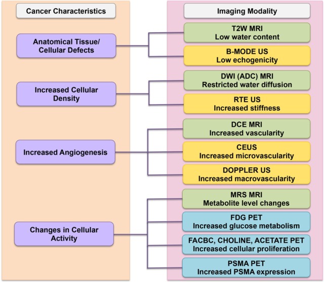
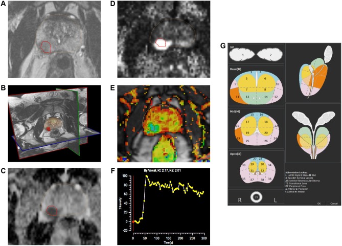
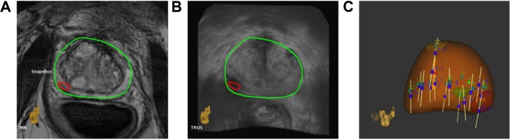
Imaging is playing an increasingly important role in the detection of prostate cancer (PCa). This review summarizes the key imaging modalities-multiparametric ultrasound (US), multiparametric magnetic resonance imaging (MRI), MRI-US fusion imaging, and positron emission tomography (PET) imaging-used in the diagnosis and localization of PCa. Emphasis is laid on the biological and functional characteristics of tumors that rationalize the use of a specific imaging technique. Changes to anatomical architecture of tissue can be detected by anatomical grayscale US and T2-weighted MRI. Tumors are known to progress through angiogenesis-a fact exploited by Doppler and contrast-enhanced US and dynamic contrast-enhanced MRI. The increased cellular density of tumors is targeted by elastography and diffusion-weighted MRI. PET imaging employs several different radionuclides to target the metabolic and cellular activities during tumor growth. Results from studies using these various imaging techniques are discussed and compared.
成像在前列腺癌(PCa)的检测中发挥着越来越重要的作用。本综述总结了用于PCa诊断和定位的关键成像模态——多参数超声(US)、多参数磁共振成像(MRI)、MRI-US融合成像和正电子发射断层扫描(PET)成像。重点阐述了使特定成像技术得以合理应用的肿瘤生物学和功能特征。组织解剖结构的变化可通过解剖灰阶超声和T2加权MRI检测到。已知肿瘤通过血管生成进展——这一事实被多普勒超声、超声造影以及动态对比增强MRI所利用。弹性成像和扩散加权MRI针对肿瘤增加的细胞密度。PET成像采用几种不同的放射性核素靶向肿瘤生长过程中的代谢和细胞活动。讨论并比较了使用这些不同成像技术的研究结果。