Vetuschi A, D'Alfonso A, Sferra R, Zanelli D, Pompili S, Patacchiola F, Gaudio E, Carta G
University of L'Aquila.
Eur J Histochem. 2016 Feb 16;60(1):2604. doi: 10.4081/ejh.2016.2604.
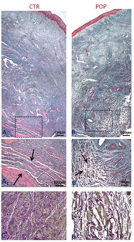
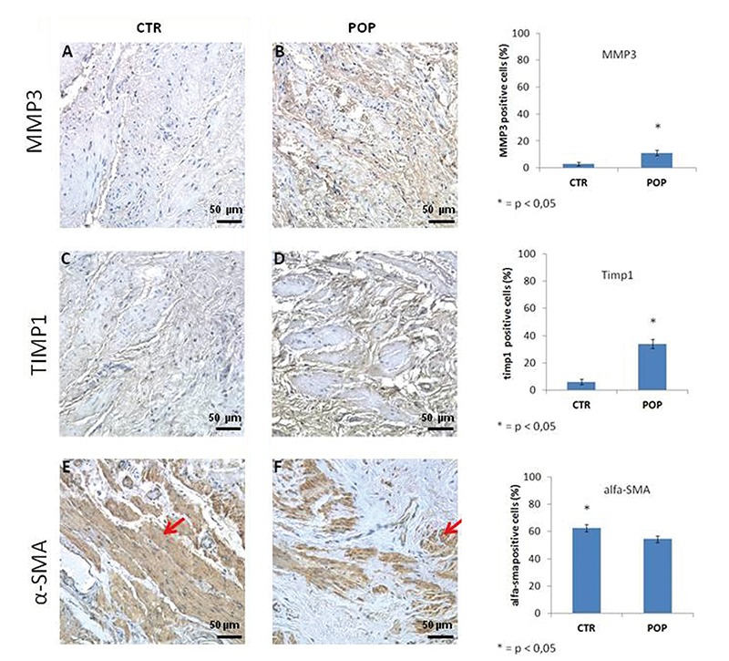
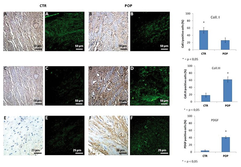
The objective of this study was to evaluate the morphological and immunohistochemical alterations of tissue removed from the upper third of anterior vaginal wall in a sample group of the female population presenting homogenous risk factors associated with Pelvic Organ Prolapse (POP). The case study consisted of 14 patients with POP and there were 10 patients in the control group. Patient selection was carried on the basis of specific criteria and all of the patients involved in the study presented one or more of the recognized POP risk factors. Samples were taken from POP patients during vaginal plastic surgery following colpohysterectomy, and from control patients during closure of the posterior fornix following hysterectomy. Samples were processed for histological and immunohistochemical analyses for Collagen I and Collagen III, α-Smooth Muscle Actin (α-SMA), Platelet-Derived-Growth-Factor (PDGF), matrix metalloproteinase 3 (MMP3), Caspase3. Immunofluorescence analyses for Collagen I and III and PDGF were also carried out. In prolapsed specimens our results show a disorganization of smooth muscle cells that appeared to have been displaced by an increased collagen III deposition resulting in rearrangement of the muscularis propria architecture. These findings suggest that the increase in the expression of collagen fibers in muscularis could probably due to a phenotypic switch resulting in the dedifferentiation of smooth muscle cells into myofibroblasts. These alterations could be responsible for the compromising of the dynamic functionality of the pelvic floor.
本研究的目的是评估在一组具有与盆腔器官脱垂(POP)相关的同质风险因素的女性人群样本中,取自阴道前壁上三分之一组织的形态学和免疫组化改变。该病例研究包括14例POP患者,对照组有10例患者。患者选择基于特定标准,参与研究的所有患者均呈现一种或多种公认的POP风险因素。样本取自POP患者在阴道子宫切除术后的阴道整形手术期间,以及对照组患者在子宫切除术后后穹窿封闭期间。样本进行了组织学和免疫组化分析,检测I型胶原和III型胶原、α-平滑肌肌动蛋白(α-SMA)、血小板衍生生长因子(PDGF)、基质金属蛋白酶3(MMP3)、半胱天冬酶3。还进行了I型胶原和III型胶原以及PDGF的免疫荧光分析。在脱垂标本中,我们的结果显示平滑肌细胞紊乱,似乎被III型胶原沉积增加所取代,导致固有肌层结构重新排列。这些发现表明,肌层中胶原纤维表达的增加可能是由于表型转换导致平滑肌细胞去分化为肌成纤维细胞。这些改变可能是盆底动态功能受损的原因。