El-Sharkawy Mohamed, El-Mazny Akmal, Ramadan Wafaa, Hatem Dina, Abdel-Hafiz Aly, Hammam Mohamed, Nada Adel
Department of Obstetrics and Gynecology, Faculty of Medicine, Cairo University, Cairo, Egypt.
BMC Womens Health. 2016 Mar 16;16:18. doi: 10.1186/s12905-016-0297-3.
Ultrasonography has been extensively used in women suspected of having a gynecological malignancy. The aim of this study is to evaluate the efficacy of 3D ultrasonography and power Doppler for discrimination between benign and malignant endometrium in premenopausal women with abnormal uterine bleeding.
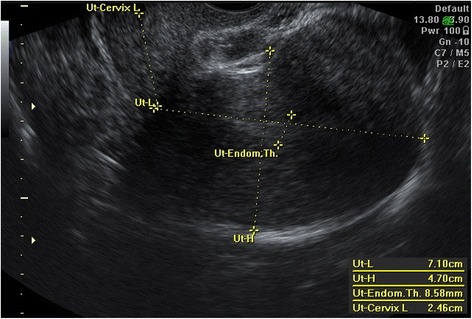
This cross-sectional study included 78 premenopausal women with abnormal uterine bleeding scheduled for hysteroscopy and endometrial curettage. The endometrial thickness (ET), uterine artery pulsatility index (PI) and resistance index (RI), and endometrial volume (EV) and 3D power Doppler vascularization index (VI), flow index (FI), and vascularization flow index (VFI) were measured and compared with hysteroscopic and histopathologic findings.
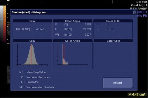
The ET (P <0.001), EV (P <0.001), and endometrial VI (P <0.001) and VFI (P = 0.043) were significantly increased in patients with atypical endometrial hyperplasia and endometrial carcinoma (n = 10) than those with benign endometrium (n = 68); whereas, the uterine artery PI and RI and endometrial FI were not significantly different between the two groups. The best marker for discrimination between benign and malignant endometrium was the VI with an area under the ROC curve of 0.88 at a cutoff value of 0.81%.
3D ultrasonography and power Doppler, especially endometrial VI, may be useful for discrimination between benign and malignant endometrium in premenopausal women with abnormal uterine bleeding.
超声检查已广泛应用于疑似患有妇科恶性肿瘤的女性。本研究的目的是评估三维超声和能量多普勒在鉴别绝经前异常子宫出血女性的良性和恶性子宫内膜方面的效能。
这项横断面研究纳入了78例计划进行宫腔镜检查和子宫内膜刮宫术的绝经前异常子宫出血女性。测量子宫内膜厚度(ET)、子宫动脉搏动指数(PI)和阻力指数(RI),以及子宫内膜体积(EV)和三维能量多普勒血管化指数(VI)、血流指数(FI)和血管化血流指数(VFI),并与宫腔镜检查和组织病理学结果进行比较。
非典型子宫内膜增生和子宫内膜癌患者(n = 10)的ET(P <0.001)、EV(P <0.001)、子宫内膜VI(P <0.001)和VFI(P = 0.043)显著高于良性子宫内膜患者(n = 68);而两组之间子宫动脉PI、RI和子宫内膜FI无显著差异。鉴别良性和恶性子宫内膜的最佳标志物是VI,在截断值为0.81%时,ROC曲线下面积为0.88。
三维超声和能量多普勒,尤其是子宫内膜VI,可能有助于鉴别绝经前异常子宫出血女性的良性和恶性子宫内膜。