Kim Chae-Lim, Cha Sun-Yeong, Chun Min Young, Kim Bumsoo, Choi Min Young, Cheon Yong-Pil
Division of Developmental Biology and Physiology, School of Biosciences and Chemistry, Institute for Basic Sciences, Sungshin Women's University, Seoul 142-742, Korea.
Global Medical Science, Sungshin Women's University, Seoul 142-742, Korea.
Dev Reprod. 2015 Sep;19(3):145-52. doi: 10.12717/DR.2015.19.3.145.
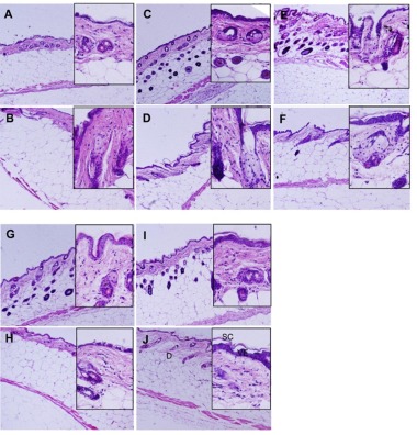
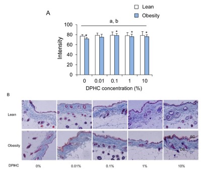
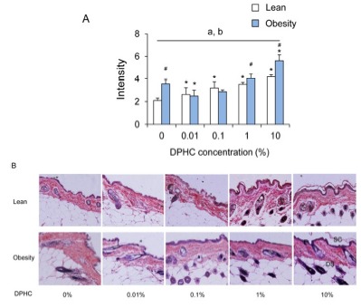
Diphlorethohydroxycarmalol (DPHC) is a known to modulate the expression of extracellular matrix (ECM) components in 3T3-L1. However, the possible role of DPHC in integument stability during obesity induction is not clear yet. We evaluated the effects of DPHC on collagen or elastic fiber quantity in integument during obesity induction with high-fat diet. The dorsal back integument sections were stained with hematoxylin-eosin, Masson trichrome, and Verhoff-Van Gieson. The intensities of collagen fibers and elastin fibers were analyzed with ImageJ. The number of fibroblasts was counted at ×1,000 fields. The number of fibroblast was increased by obesity induction, but DPHC suppressed it in a concentrationdependent manner both in lean and obese mice. On the other hand, the intensities of collagen fibers were increased by DPHC treatment in obese mice groups but not in lean mice groups. The intensities of collagen fibers of obese mice were lower than that of the lean mice in 0% group. However, the number became similar between lean and obese mice by the treatment of DPHC. The intensity of elastic fibers was increased in the lean mice with the concentration of DPHC. In the obese mice group, there were increasing patterns but only significant at 10% DPHC group. The intensity of elastic fibers of obese mice was higher than lean mice in 0%, 1%, and 10% groups. Histologically epithelial cells and follicle cells which were diffused nuclear staining forms were increased by DPHC treatment. The results suggest that the activity of integument cells during obesity induction can be modulated by DPHC.
二聚倍半萜羟基卡马醇(DPHC)已知可调节3T3-L1细胞外基质(ECM)成分的表达。然而,DPHC在肥胖诱导过程中对皮肤稳定性的可能作用尚不清楚。我们评估了DPHC对高脂饮食诱导肥胖过程中皮肤中胶原蛋白或弹性纤维数量的影响。背部皮肤切片用苏木精-伊红、Masson三色染色法和Verhoff-Van Gieson染色法染色。用ImageJ分析胶原纤维和弹性纤维的强度。在×1000视野下计数成纤维细胞的数量。肥胖诱导使成纤维细胞数量增加,但DPHC在瘦小鼠和肥胖小鼠中均以浓度依赖性方式抑制了这一增加。另一方面,DPHC处理使肥胖小鼠组的胶原纤维强度增加,但瘦小鼠组未增加。在0%组中,肥胖小鼠的胶原纤维强度低于瘦小鼠。然而,通过DPHC处理,瘦小鼠和肥胖小鼠之间的数量变得相似。随着DPHC浓度的增加,瘦小鼠的弹性纤维强度增加。在肥胖小鼠组中,有增加的趋势,但仅在10% DPHC组有显著增加。在0%、1%和10%组中,肥胖小鼠的弹性纤维强度高于瘦小鼠。组织学上,DPHC处理增加了呈弥漫性核染色形式的上皮细胞和毛囊细胞。结果表明,DPHC可以调节肥胖诱导过程中皮肤细胞的活性。