Bragança Bruno, Oliveira-Monteiro Nádia, Ferreirinha Fátima, Lima Pedro A, Faria Miguel, Fontes-Sousa Ana P, Correia-de-Sá Paulo
Laboratório de Farmacologia e Neurobiologia - Center for Drug Discovery and Innovative Medicines (MedInUP), Instituto de Ciências Biomédicas Abel Salazar (ICBAS), Universidade do Porto (UP) Porto, Portugal.
Departamento de Química e Bioquímica, Faculdade de Ciências, Centro de Química e Bioquímica, Universidade de Lisboa Lisboa, Portugal.
Front Pharmacol. 2016 Mar 7;7:45. doi: 10.3389/fphar.2016.00045. eCollection 2016.
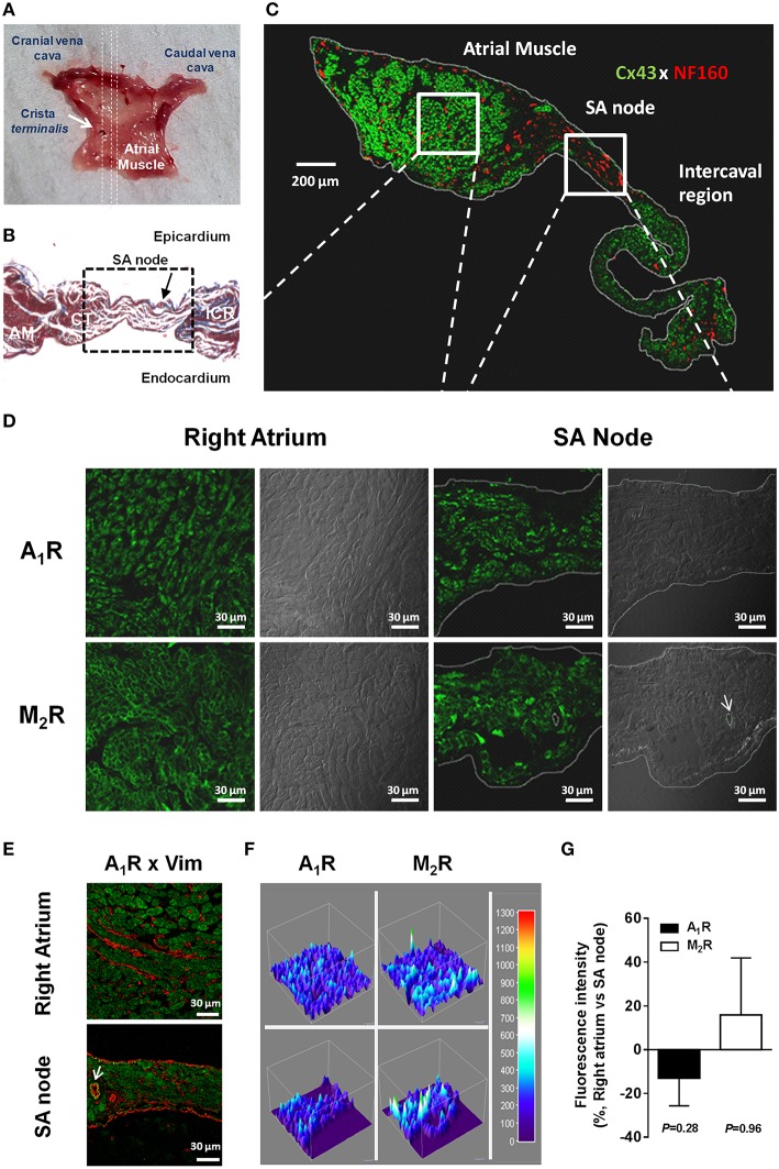
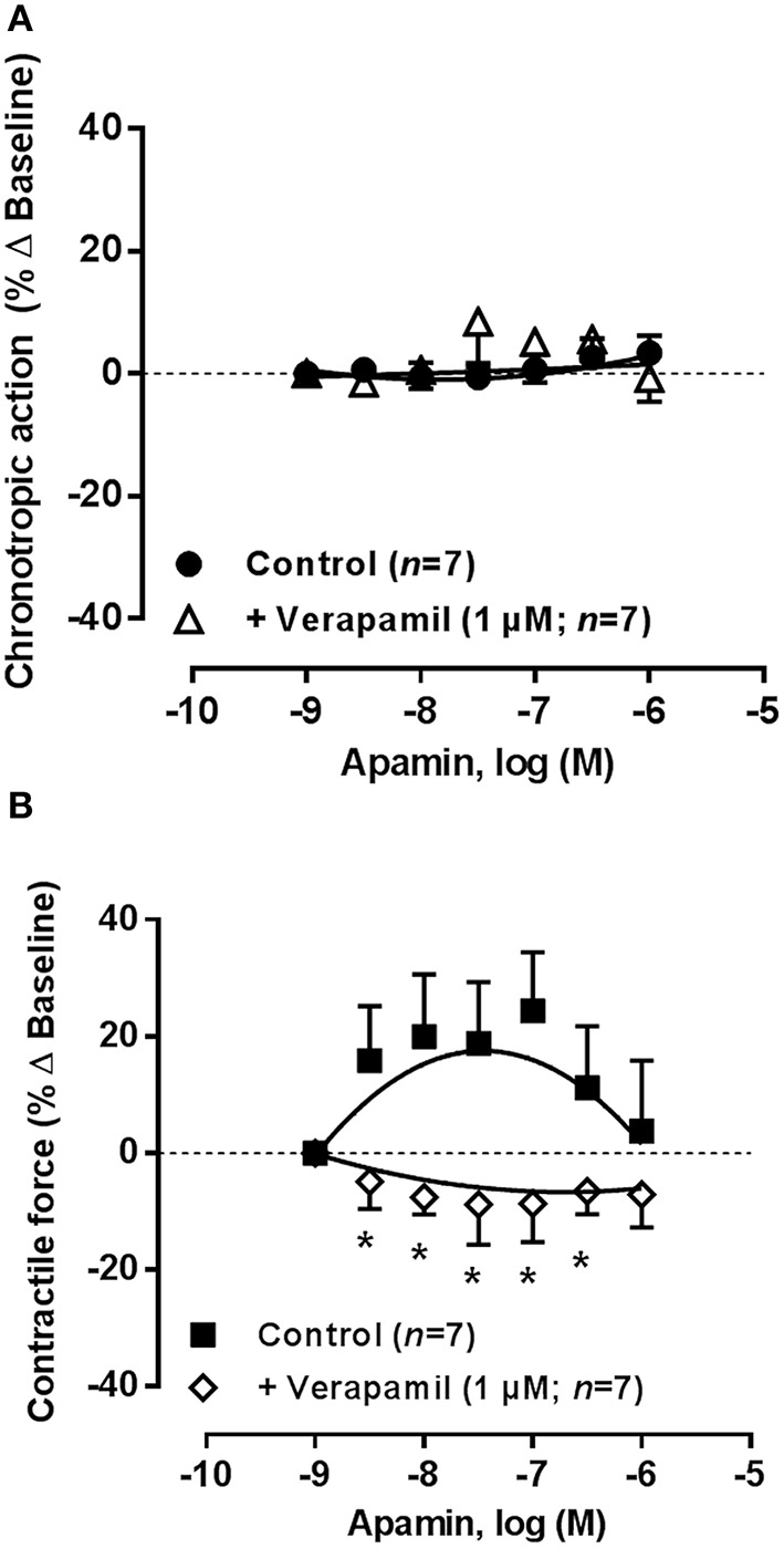
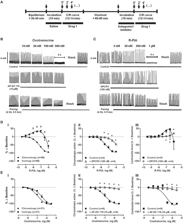
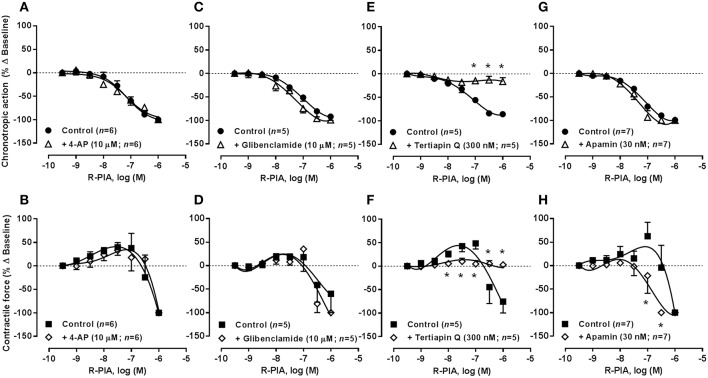
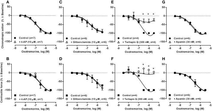
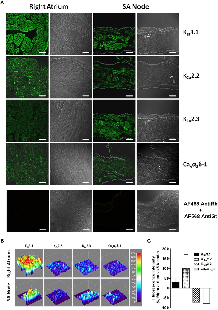
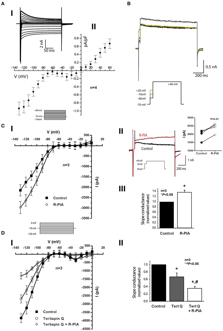
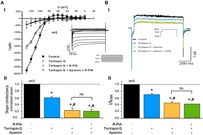
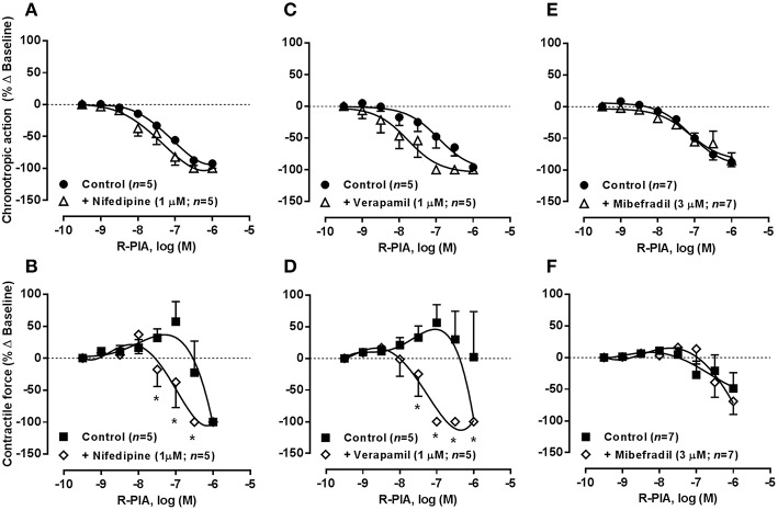
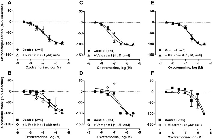
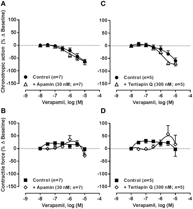
Impulse generation in supraventricular tissue is inhibited by adenosine and acetylcholine via the activation of A1 and M2 receptors coupled to inwardly rectifying GIRK/KIR3.1/3.4 channels, respectively. Unlike M2 receptors, bradycardia produced by A1 receptors activation predominates over negative inotropy. Such difference suggests that other ion currents may contribute to adenosine chronoselectivity. In isolated spontaneously beating rat atria, blockade of KCa2/SK channels with apamin and Cav1 (L-type) channels with nifedipine or verapamil, sensitized atria to the negative inotropic action of the A1 agonist, R-PIA, without affecting the nucleoside negative chronotropy. Patch-clamp experiments in the whole-cell configuration mode demonstrate that adenosine, via A1 receptors, activates the inwardly-rectifying GIRK/KIR3.1/KIR3.4 current resulting in hyperpolarization of atrial cardiomyocytes, which may slow down heart rate. Conversely, the nucleoside inactivates a small conductance Ca(2+)-activated KCa2/SK outward current, which eventually reduces the repolarizing force and thereby prolong action potentials duration and Ca(2+) influx into cardiomyocytes. Immunolocalization studies showed that differences in A1 receptors distribution between the sinoatrial node and surrounding cardiomyocytes do not afford a rationale for adenosine chronoselectivity. Immunolabelling of KIR3.1, KCa2.2, KCa2.3, and Cav1 was also observed throughout the right atrium. Functional data indicate that while both A1 and M2 receptors favor the opening of GIRK/KIR3.1/3.4 channels modulating atrial chronotropy, A1 receptors may additionally restrain KCa2/SK activation thereby compensating atrial inotropic depression by increasing the time available for Ca(2+) influx through Cav1 (L-type) channels.
腺苷和乙酰胆碱分别通过激活与内向整流GIRK/KIR3.1/3.4通道偶联的A1和M2受体,抑制室上组织中的冲动产生。与M2受体不同,A1受体激活所产生的心动过缓比负性肌力作用更为显著。这种差异表明,其他离子电流可能与腺苷的时间选择性有关。在离体自发搏动的大鼠心房中,用蜂毒明肽阻断KCa2/SK通道,用硝苯地平或维拉帕米阻断Cav1(L型)通道,可使心房对A1激动剂R-PIA的负性肌力作用敏感化,而不影响核苷的负性变时作用。全细胞膜片钳实验表明,腺苷通过A1受体激活内向整流GIRK/KIR3.1/KIR3.4电流,导致心房心肌细胞超极化,这可能会减慢心率。相反,核苷可使小电导Ca(2+)激活的KCa2/SK外向电流失活,最终降低复极力,从而延长动作电位持续时间,并增加Ca(2+)流入心肌细胞。免疫定位研究表明,窦房结和周围心肌细胞之间A1受体分布的差异并不能解释腺苷的时间选择性。在整个右心房也观察到了KIR3.1、KCa2.2、KCa2.3和Cav1的免疫标记。功能数据表明,虽然A1和M2受体都有利于GIRK/KIR3.1/3.4通道开放,从而调节心房变时性,但A1受体可能还会抑制KCa2/SK的激活,从而通过增加Ca(2+)通过Cav1(L型)通道流入的时间,来补偿心房的正性肌力抑制作用。