Crupi Rosalia, Impellizzeri Daniela, Bruschetta Giuseppe, Cordaro Marika, Paterniti Irene, Siracusa Rosalba, Cuzzocrea Salvatore, Esposito Emanuela
Department of Biological and Environmental Sciences, University of Messina Messina, Italy.
Department of Biological and Environmental Sciences, University of MessinaMessina, Italy; Manchester Biomedical Research Centre, Manchester Royal Infirmary, School of Medicine, The University of ManchesterManchester, UK.
Front Pharmacol. 2016 Mar 8;7:47. doi: 10.3389/fphar.2016.00047. eCollection 2016.
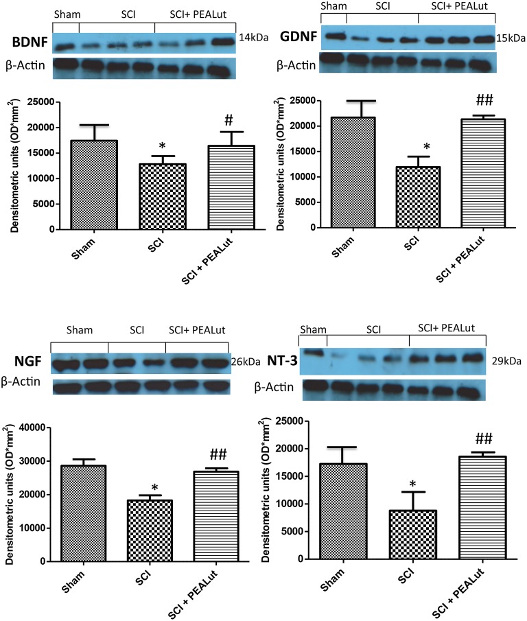
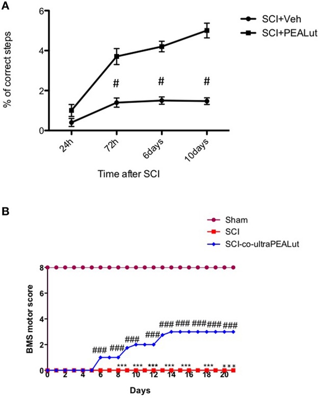
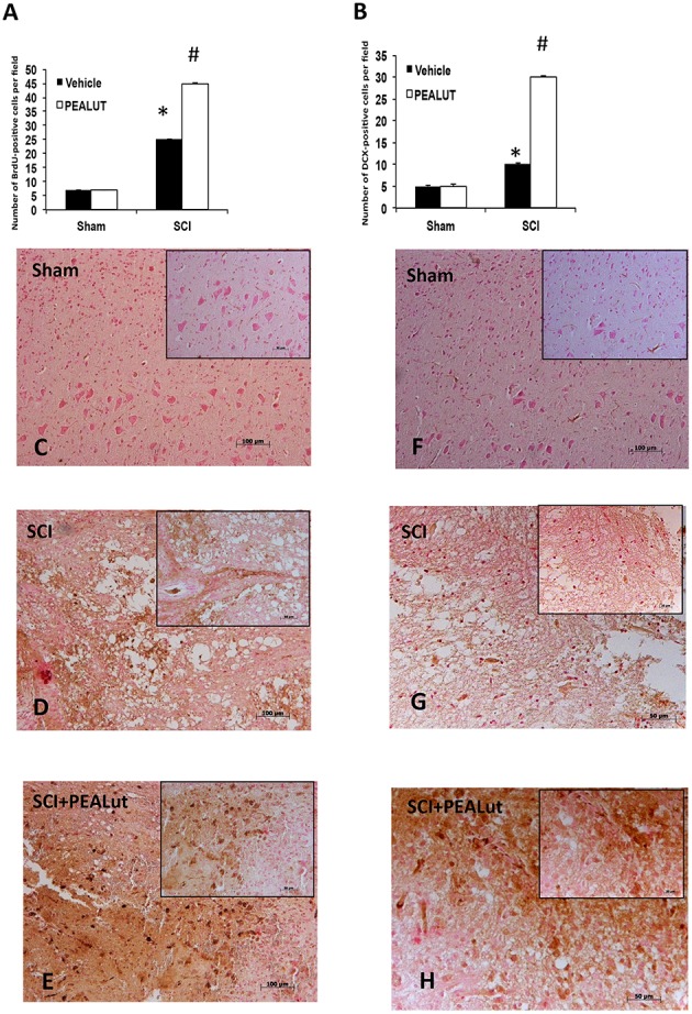
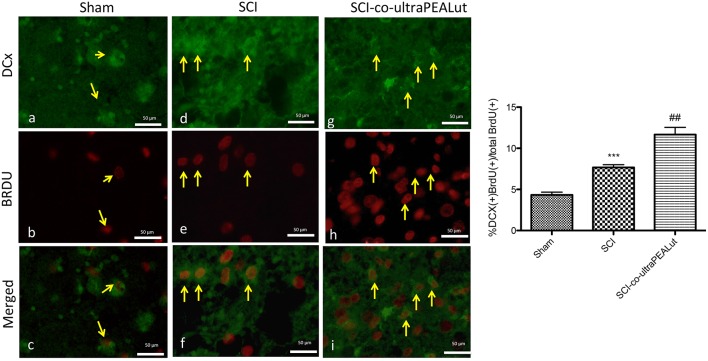
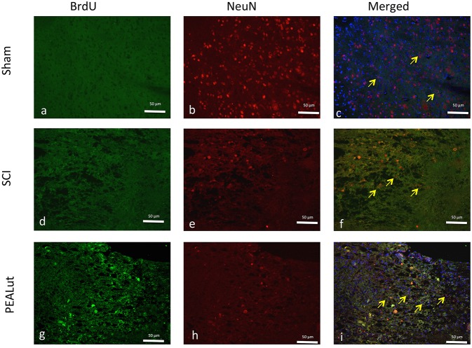
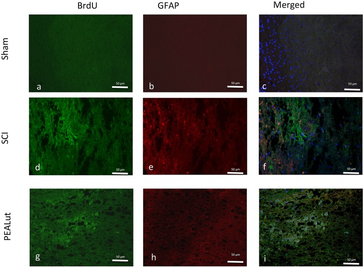
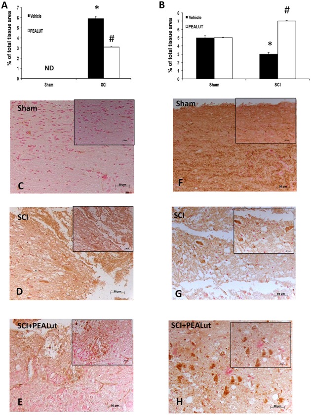
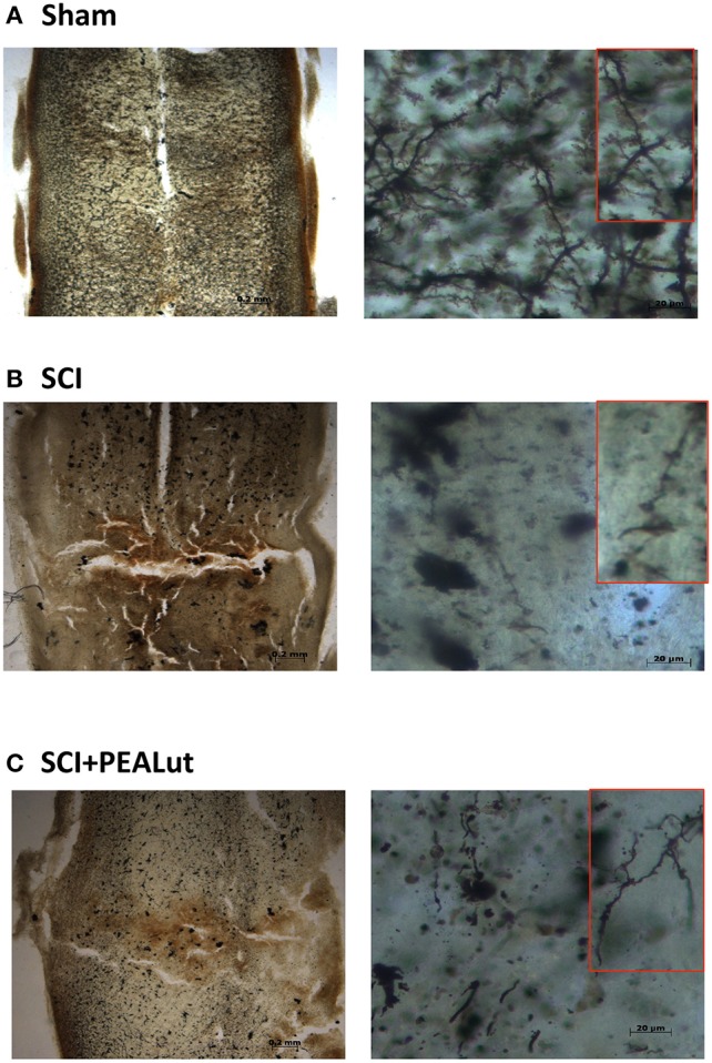
Spinal cord injury (SCI) stimulates activation of astrocytes and infiltration of immune cells at the lesion site; however, the mechanism that promotes the birth of new neurons is still under debate. Neuronal regeneration is restricted after spinal cord injury, but can be stimulated by experimental intervention. Previously we demonstrated that treatment co-ultramicronized palmitoylethanolamide and luteolin, namely co-ultraPEALut, reduced inflammation. The present study was designed to explore the neuroregenerative properties of co-ultraPEALut in an estabished murine model of SCI. A vascular clip was applied to the spinal cord dura at T5-T8 to provoke injury. Mice were treated with co-ultraPEALut (1 mg/kg, intraperitoneally) daily for 72 h after SCI. Co-ultraPEALut increased the numbers of both bromodeoxyuridine-positive nuclei and doublecortin-immunoreactive cells in the spinal cord of injured mice. To correlate neuronal development with synaptic plasticity a Golgi method was employed to analyze dendritic spine density. Co-ultraPEALut administration stimulated expression of the neurotrophic factors brain-derived neurotrophic factor, glial cell-derived neurotrophic factor, nerve growth factor, and neurotrophin-3. These findings show a prominent effect of co-ultraPEALut administration in the management of survival and differentiation of new neurons and spine maturation, and may represent a therapeutic treatment for spinal cord and other traumatic diseases.
脊髓损伤(SCI)会刺激损伤部位星形胶质细胞的活化和免疫细胞的浸润;然而,促进新神经元生成的机制仍存在争议。脊髓损伤后神经元再生受到限制,但可通过实验干预来刺激。此前我们证明,联合超微化棕榈酰乙醇胺和木犀草素(即联合超微化PEA-Lut)的治疗可减轻炎症。本研究旨在探讨联合超微化PEA-Lut在已建立的小鼠脊髓损伤模型中的神经再生特性。在T5-T8水平对脊髓硬脊膜应用血管夹以引发损伤。脊髓损伤后,小鼠每天腹腔注射联合超微化PEA-Lut(1 mg/kg),持续72小时。联合超微化PEA-Lut增加了损伤小鼠脊髓中溴脱氧尿苷阳性细胞核和双皮质素免疫反应性细胞的数量。为了将神经元发育与突触可塑性相关联,采用高尔基方法分析树突棘密度。联合超微化PEA-Lut给药刺激了神经营养因子脑源性神经营养因子、胶质细胞源性神经营养因子、神经生长因子和神经营养素-3的表达。这些发现表明联合超微化PEA-Lut给药在新神经元的存活和分化以及树突棘成熟的管理方面具有显著作用,可能代表了一种治疗脊髓和其他创伤性疾病的方法。