Lashkari AmirEhsan, Pak Fatemeh, Firouzmand Mohammad
Department of Bio-Medical Engineering, Institute of Electrical Engineering and Information Technology, Iranian Research Organization for Science and Technology, Tehran, Iran.
J Med Signals Sens. 2016 Jan-Mar;6(1):12-24.
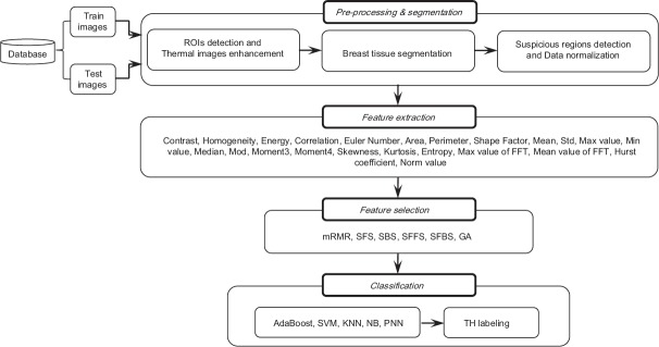
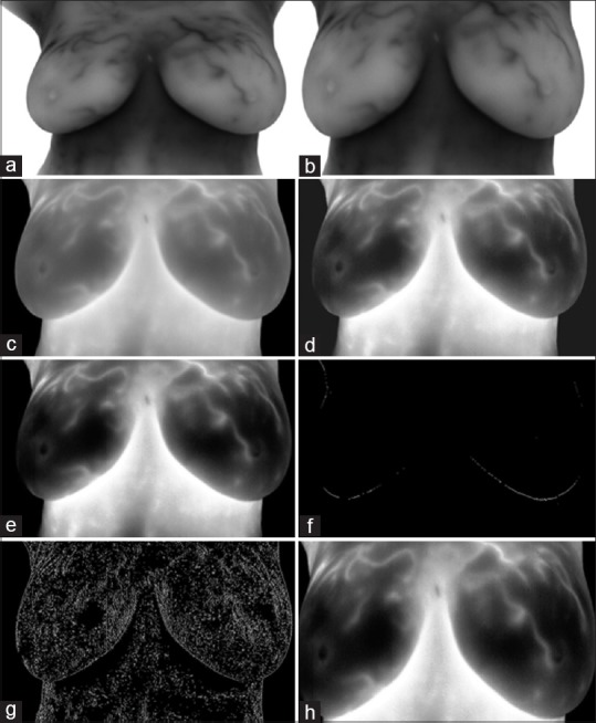
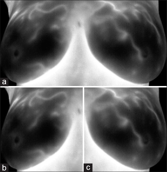
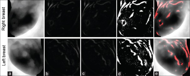
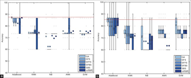
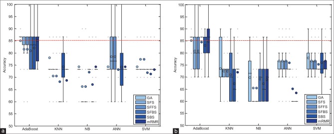
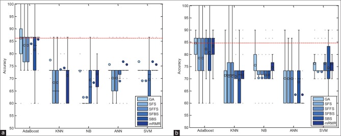
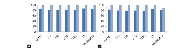
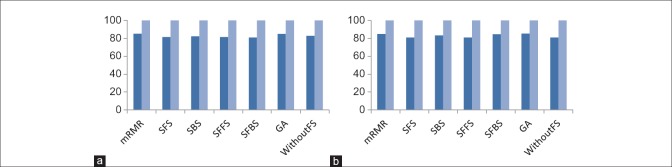
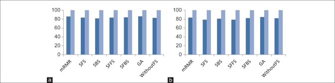
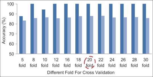
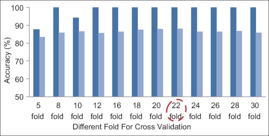
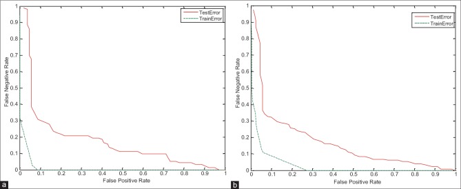
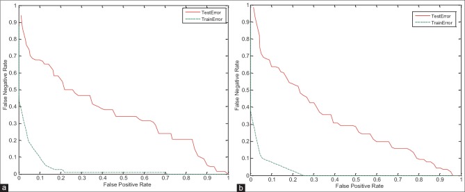
Breast cancer is the most common type of cancer among women. The important key to treat the breast cancer is early detection of it because according to many pathological studies more than 75% - 80% of all abnormalities are still benign at primary stages; so in recent years, many studies and extensive research done to early detection of breast cancer with higher precision and accuracy. Infra-red breast thermography is an imaging technique based on recording temperature distribution patterns of breast tissue. Compared with breast mammography technique, thermography is more suitable technique because it is noninvasive, non-contact, passive and free ionizing radiation. In this paper, a full automatic high accuracy technique for classification of suspicious areas in thermogram images with the aim of assisting physicians in early detection of breast cancer has been presented. Proposed algorithm consists of four main steps: pre-processing & segmentation, feature extraction, feature selection and classification. At the first step, using full automatic operation, region of interest (ROI) determined and the quality of image improved. Using thresholding and edge detection techniques, both right and left breasts separated from each other. Then relative suspected areas become segmented and image matrix normalized due to the uniqueness of each person's body temperature. At feature extraction stage, 23 features, including statistical, morphological, frequency domain, histogram and Gray Level Co-occurrence Matrix (GLCM) based features are extracted from segmented right and left breast obtained from step 1. To achieve the best features, feature selection methods such as minimum Redundancy and Maximum Relevance (mRMR), Sequential Forward Selection (SFS), Sequential Backward Selection (SBS), Sequential Floating Forward Selection (SFFS), Sequential Floating Backward Selection (SFBS) and Genetic Algorithm (GA) have been used at step 3. Finally to classify and TH labeling procedures, different classifiers such as AdaBoost, Support Vector Machine (SVM), k-Nearest Neighbors (kNN), Naïve Bayes (NB) and probability Neural Network (PNN) are assessed to find the best suitable one. These steps are applied on different thermogram images degrees. The results obtained on native database showed the best and significant performance of the proposed algorithm in comprise to the similar studies. According to experimental results, GA combined with AdaBoost with the mean accuracy of 85.33% and 87.42% on the left and right breast images with 0 degree, GA combined with AdaBoost with mean accuracy of 85.17% on the left breast images with 45 degree and mRMR combined with AdaBoost with mean accuracy of 85.15% on the right breast images with 45 degree, and also GA combined with AdaBoost with a mean accuracy of 84.67% and 86.21%, on the left and right breast images with 90 degree, are the best combinations of feature selection and classifier for evaluation of breast images.
乳腺癌是女性中最常见的癌症类型。治疗乳腺癌的关键在于早期发现,因为根据许多病理学研究,在所有异常情况中,超过75% - 80%在初级阶段仍为良性;因此,近年来开展了许多研究和广泛的探索,以更高的精度和准确性对乳腺癌进行早期检测。红外乳腺热成像术是一种基于记录乳腺组织温度分布模式的成像技术。与乳腺钼靶摄影技术相比,热成像术更具优势,因为它是非侵入性、非接触式、被动式且无电离辐射。本文提出了一种全自动高精度技术,用于对热成像图中的可疑区域进行分类,旨在协助医生早期发现乳腺癌。所提算法包括四个主要步骤:预处理与分割、特征提取、特征选择和分类。第一步,通过全自动操作确定感兴趣区域(ROI)并改善图像质量。利用阈值化和边缘检测技术,将左右乳房彼此分离。然后,由于每个人体温的独特性,相对可疑区域被分割出来,图像矩阵被归一化。在特征提取阶段,从第一步获得的分割后的左右乳房中提取23个特征,包括基于统计、形态学、频域、直方图和灰度共生矩阵(GLCM)的特征。为了获得最佳特征,在第三步使用了诸如最小冗余最大相关性(mRMR)、顺序前向选择(SFS)、顺序后向选择(SBS)、顺序浮动前向选择(SFFS)、顺序浮动后向选择(SFBS)和遗传算法(GA)等特征选择方法。最后,为了进行分类和TH标记程序,评估了不同的分类器,如AdaBoost、支持向量机(SVM)、k近邻(kNN)、朴素贝叶斯(NB)和概率神经网络(PNN),以找到最合适的分类器。这些步骤应用于不同程度的热成像图。在本地数据库上获得的结果表明,与类似研究相比,所提算法具有最佳且显著的性能。根据实验结果,GA与AdaBoost相结合,在0度的左右乳房图像上平均准确率分别为85.33%和87.42%;GA与AdaBoost相结合,在45度的左乳房图像上平均准确率为85.17%;mRMR与AdaBoost相结合,在45度的右乳房图像上平均准确率为85.15%;GA与AdaBoost相结合,在90度的左右乳房图像上平均准确率分别为84.67%和86.21%,这些是用于评估乳房图像的特征选择和分类器的最佳组合。