Milosevic Marina, Jankovic Dragan, Peulic Aleksandar
Department of Computer Engineering, Faculty of Technical Sciences, University of Kragujevac, Serbia.
Department of Computer Science, Faculty of Electronic Engineering, University of Nis, Serbia.
EXCLI J. 2014 Nov 4;13:1204-15. eCollection 2014.
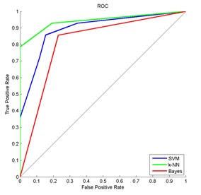
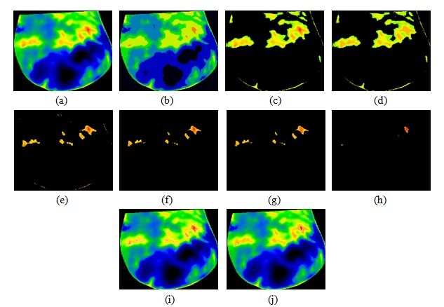
In this paper, we present a system based on feature extraction techniques and image segmentation techniques for detecting and diagnosing abnormal patterns in breast thermograms. The proposed system consists of three major steps: feature extraction, classification into normal and abnormal pattern and segmentation of abnormal pattern. Computed features based on gray-level co-occurrence matrices are used to evaluate the effectiveness of textural information possessed by mass regions. A total of 20 GLCM features are extracted from thermograms. The ability of feature set in differentiating abnormal from normal tissue is investigated using a Support Vector Machine classifier, Naive Bayes classifier and K-Nearest Neighbor classifier. To evaluate the classification performance, five-fold cross validation method and Receiver operating characteristic analysis was performed. The verification results show that the proposed algorithm gives the best classification results using K-Nearest Neighbor classifier and a accuracy of 92.5%. Image segmentation techniques can play an important role to segment and extract suspected hot regions of interests in the breast infrared images. Three image segmentation techniques: minimum variance quantization, dilation of image and erosion of image are discussed. The hottest regions of thermal breast images are extracted and compared to the original images. According to the results, the proposed method has potential to extract almost exact shape of tumors.
在本文中,我们提出了一种基于特征提取技术和图像分割技术的系统,用于检测和诊断乳腺热图中的异常模式。所提出的系统包括三个主要步骤:特征提取、分类为正常和异常模式以及异常模式的分割。基于灰度共生矩阵计算的特征用于评估肿块区域所具有的纹理信息的有效性。从热图中总共提取了20个灰度共生矩阵特征。使用支持向量机分类器、朴素贝叶斯分类器和K近邻分类器研究了特征集区分异常组织和正常组织的能力。为了评估分类性能,进行了五折交叉验证方法和接收器操作特性分析。验证结果表明,所提出的算法使用K近邻分类器给出了最佳分类结果,准确率为92.5%。图像分割技术在分割和提取乳腺红外图像中可疑的感兴趣热区方面可以发挥重要作用。讨论了三种图像分割技术:最小方差量化、图像膨胀和图像腐蚀。提取了乳腺热图像中最热的区域并与原始图像进行比较。根据结果,所提出的方法有潜力提取几乎精确的肿瘤形状。